
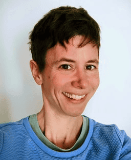
Simone de Brot is a veterinary pathologist at the Institute of Animal Pathology of the Department of Infectious Diseases and Pathobiology (DIP) of the University of Bern, Switzerland. The DIP carries out biomedical research to improve health and well-being of animals and humans. It is strongly involved in teaching and education in veterinary medicine, and includes not only students of veterinary medicine, but also those of medicine and natural sciences. The DIP offers research-supported diagnostic services to various stakeholders, including academic and private clinics, veterinary public health institutions and medical laboratories and clinicians.
Please tell us a bit about your research center and what you do.
At the Institute of Animal Pathology, University of Bern, we examine tissues from a wide variety of animal species for diagnostic, teaching, and research purposes. Our institute’s own research primarily focuses on cancer and host–pathogen interactions. Within our core facility COMPATH (Comparative Pathology), a joint University of Bern platform for human and veterinary pathology, we mainly analyze tissues from animal models as well as in vitro models provided by internal and external research groups from academia, industry, and the private sector. Our work typically involves the evaluation of histological sections, often in combination with immunohistochemistry or immunofluorescence, to address specific scientific questions. These analyses are largely performed using digital pathology approaches. Digital workflows allow us to ensure objective, standardized, and reproducible evaluation of tissue sections. This approach also facilitates quantitative image analysis and supports collaborative research projects.
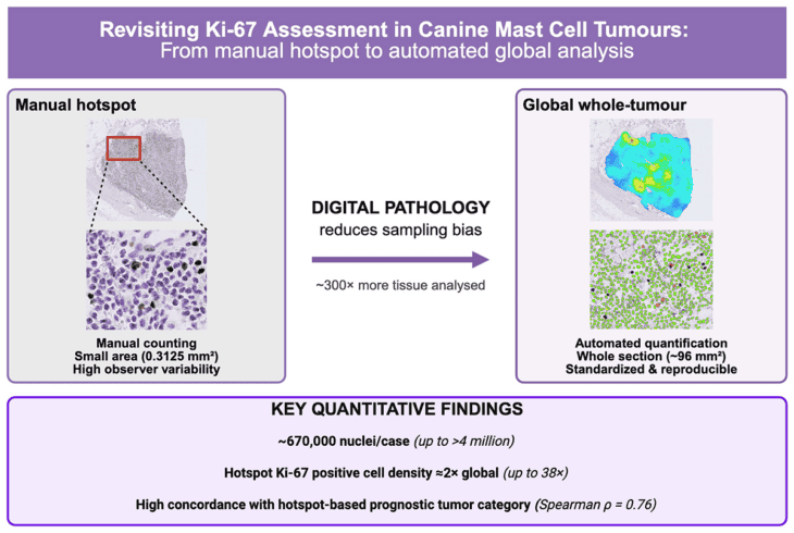
Graphical abstract for: Revisiting Ki-67 Assessment in Canine Mast Cell Tumours: From Manual Hotspot to Automated Global Analysis, DOI: 10.3390/vetsci13020198
Which kind of problems or questions do you address with Visiopharm? How did Visiopharm help you overcome those initial challenges?
The size of our projects is highly variable. We frequently handle small to medium-sized projects, often consisting of only 10 to 100 tissue sections. At the same time, these samples show considerable variability with regard to tissue type, extent and nature of lesions, as well as tissue and staining quality. As a result, predefined digital analysis solutions, often developed for human and mouse tissues using standardized staining protocols, are frequently not applicable in our setting. With only a few exceptions, we therefore develop project-specific analytical applications (APPs) tailored to the individual study. To keep the required effort manageable and enable pathologists themselves to perform these analyses, we rely on user-friendly software. Equally important are integrated options for deep learning classifiers, a wide range of post-processing steps, and flexible output formats. In the vast majority of cases, Visiopharm provides the solution that allows us to address these diverse analytical challenges efficiently.
“In our work with highly heterogeneous samples across many animal species and study designs, flexibility is essential. Visiopharm gives us the freedom to build tailored analyses while remaining intuitive enough for pathologists to use efficiently.“
Simone de Brot
What specific capabilities of the product do you find most helpful?
A particularly useful feature for our work is the ability to combine deep learning and threshold classifiers within a single app. We frequently use this approach in a two-step workflow. In a first step, a deep learning classifier is used to identify the specific cell type or tissue structure that should be evaluated for a selected tissue marker (for example, goblet cells in the small intestine). In a second step, threshold-based classification is applied to categorize these cells based on staining characteristics, such as distinguishing neutral from acidic goblet cells.
Another highly specialized but important function for us is the ability to define an object (e.g., a cell nucleus) as belonging to a category (e.g., positive for an IHC marker) based on selected central pixel values. For instance, we may analyze only the lowest 5% of pixel intensities to detect small punctate signals, such as those observed in in situ hybridization. Conversely, for more diffusely expressed markers, we may evaluate the central 25–75% of pixel intensities, which helps exclude edge artifacts or small nonspecific staining patterns.
Finally, a feature that greatly improves our time management and overall workflow across nearly all applications is batch processing. This allows analyses to be performed in the background without interrupting other ongoing work.
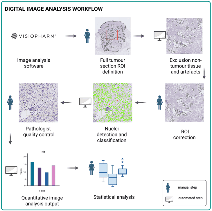
Comprehensive workflow for digital Ki-67 quantification in tumour tissue sections; from: DOI: 10.3390/vetsci13020198
Could you share some insights into how the software has added value to your project if any ? How does it make your work easier?
Digital analyses using Visiopharm software have fundamentally transformed our work as veterinary pathologists in the evaluation of tissue sections from scientific studies, very much for the better. Traditional semi-quantitative assessments, which were often imprecise and somewhat subjective, have largely been replaced by precise, fully quantitative, and clearly reproducible analyses. As a result, we can extract substantially more information from each tissue section, greatly improving the quality of microscopy-based studies.
Researchers have recognized these possibilities, and we are experiencing a strong and growing demand for such analyses at our institute. At the same time, the software provides an excellent tool to directly compare traditional histological evaluations with digital approaches, allowing us to actively contribute to the development and implementation of digital pathology. This is particularly important in veterinary medicine, where digital pathology is still less widely adopted and offers considerable potential for future growth.
Importantly, what has become easier is not necessarily the analyses themselves, these have actually become more sophisticated and complex. Rather, our work is now better supported by robust digital tools. Communication and confidence in the performed histological analyses have improved substantially, making it easier to present, discuss, and justify our findings in scientific collaborations.
Examples of their published work:
Deep Learning-based Whole-Slide Ki-67 Analysis in Canine Mast Cell Tumors: Correlation with Traditional Scoring Systems and KIT Mutation Status.
Dietrich N, Klopfleisch R, Conrad T, Puget C, Rottenberg S, Kiupel M, de Brot S, Under review (Pathology Informatics)
Revisiting Ki-67 Assessment in Canine Mast Cell Tumours: From Manual Hotspot to Automated Global Analysis.
Scalco R, Wasmer E, Jäger K, Rottenberg S, Aupperle-Lellbach H, de Brot S. , Vet Sci. 2026 Feb 18;13(2):198. Link
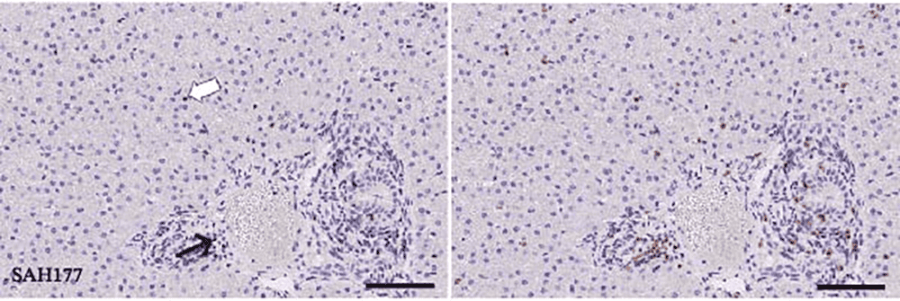
Wesselsbron Virus-Induced Hepatitis in Ewes and Lambs Unraveled Through Machine Learning-Driven Digital Histopathology.
Grau-Roma L, de Brot S, Zimoch M, Clerc L, Donzé N, Liniger M, Brito F, Herrera A, Godel A, Summerfield A, Benarafa C, García-Nicolás O., Transbound Emerg Dis. 2026 Feb 11;2026:7912840. Link
Comparative Digital Estrogen Receptor Alpha (ERα) Expression Analysis in Benign and Malignant Prostate Tissue of Men and Dogs.
Lothion-Roy J, Aeschlimann L, Hiller LA, Rottenberg S, Mongan NP, Rutland CS, Rakha E, Dean A, Rubin MA, de Brot S., Prostate. 2026 Apr;86(5):568-581. Link
 Visiopharm
Categories: Blog, Customer Stories Tags: veterinary
28900
Tech Talk #1: Faster AI Training, Tissue Alignment, and more in Visiopharm platform
Visiopharm
Categories: Blog, Customer Stories Tags: veterinary
28900
Tech Talk #1: Faster AI Training, Tissue Alignment, and more in Visiopharm platform
The first episode of the Visiopharm Tech Talks webinar series focused on helping users get more from the platform through new software capabilities, practical tips, and a preview of what’s coming next.
Hosted by Visiopharm expert, Dan Winkowski, PhD, the session highlighted recent innovations designed to make AI training faster, streamline annotation workflows, and help users get to results more efficiently.
Watch the recording of the session, or read the highlights below.
Faster and More Flexible AI Training
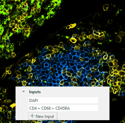
One of the key updates discussed was a series of improvements to AI training. According to Dan, recent platform enhancements have increased training speeds by up to 5x, helping users move from model development to production-ready workflows much faster.
The software now also supports combining multiple channels as inputs to AI models, including fluorescence channels and color deconvolutions. This allows users to provide richer information during training, helping create more robust and generalizable AI models.
New Annotation Experience
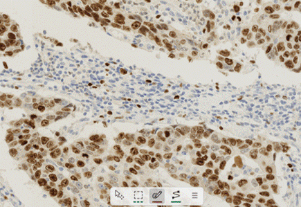
Dan highlighted the redesigned drawing tool as one of the most impactful updates in the latest release.
The new toolbar replaces the previous drawing wheel with a more intuitive, always-visible interface that supports keyboard shortcuts and works in both light and dark modes. With a smaller footprint in the viewer, it stays out of the way while making annotations faster and easier.
This is particularly useful when creating training data for AI models or selecting regions for downstream analysis.
Using Tissue Align to Connect Different Modalities
The webinar also showcased Tissue Align, Visiopharm’s multimodal image alignment technology.
Users can align images from different staining modalities, including H&E and multiplex immunofluorescence (IF), and either keep them as aligned layers or fuse them into a single dataset. This allows researchers to combine morphological information from H&E with molecular information from fluorescence markers.
Dan demonstrated how aligned datasets can help transfer annotations between image types, enabling what he described as a highly flexible, bidirectional workflow. For example, pathologists can annotate on H&E images and transfer those annotations to fluorescence data, or researchers can use fluorescence markers to guide annotations on H&E sections.
Building Better Training Data with Transfer Learning
A key theme throughout the session was improving confidence in AI training data.
Dan showed how users can identify specific cell types using marker-based stains, transfer those annotations to H&E images, and use the resulting labels to train AI models. For example, macrophages identified through CD68 staining can become training data for models that learn to recognize those same cells on H&E alone.
This approach helps users create high-quality training datasets while reducing the challenge of manually annotating difficult-to-identify cell populations.
Quick Start Apps for Faster Results
For users new to the platform, Dan strongly recommended Visiopharm’s Quick Start Apps.
These pre-trained applications are designed around common image analysis tasks and can deliver results immediately without requiring users to build workflows from scratch. The apps can also be retrained and customized with project-specific data when needed.
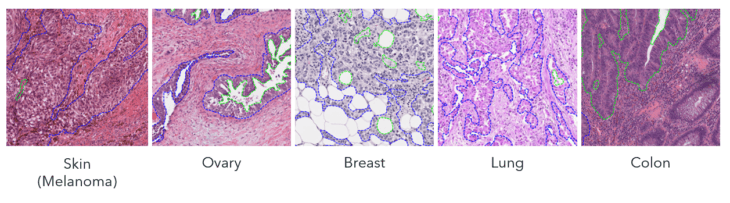
Among the latest additions is the H&E Tumor Detection Quick Start App, which has been trained across a broad range of tissues and staining conditions. The goal is simple: help users start generating meaningful results right away.
Dan also demonstrated how Quick Start Apps can significantly reduce annotation time by automatically identifying objects such as nuclei and providing a starting point for further AI training.
A Look Ahead
The session closed with a preview of a feature currently in development: enhanced clustering and visualization tools for unsupervised analysis.
The upcoming functionality will include hierarchical clustering with an interactive dendrogram, heat maps, and gallery views designed to help users explore complex datasets, including protein and transcript data at the single-cell level.
Key Takeaway
From faster AI training and improved annotation tools to multimodal image analysis and Quick Start Apps, the first Tech Talks session focused on one clear goal: helping researchers work more efficiently and get to meaningful results faster.
As Dan concluded, these updates are designed to make it easier for users to develop workflows, train AI models, and generate data with greater speed and confidence.
Sign up to the next Tech Talk session here.
Visiopharm Categories: Blog, Product Releases Tags: Tech Talk

