As we enter October, the breast cancer awareness month, we had the privilege of sitting down with Dr. Jan Trøst Jørgensen to discuss the 25th anniversary of HER2 targeted therapy – a revolutionary approach that has transformed the landscape of breast cancer treatment.
Jørgensen holds a master’s degree in pharmaceutical science and a Ph.D. in clinical pharmacy from the University of Copenhagen. With 40 years of experience in research and development at various pharmaceutical, biotech, and diagnostic companies, including Novartis, Novo Nordisk, and Dako/Agilent, he has acquired extensive expertise in the field. Currently, he serves as the Director of the Dx-Rx Institute in Fredensborg, Denmark. Dr. Jørgensen is a strong advocate for individualized pharmacotherapy and has authored and edited numerous scientific papers on companion diagnostics, drug-diagnostic co-development, and precision medicine. Furthermore, he serves as an editorial board member for several medical journals and is a board member of the Danish Society of Cyto- and Histochemistry.
Dive into our enlightening conversation as we explore this milestone and its lasting impact on patient care.
Visiopharm: Welcome Jan, can you tell us a little bit about you and your background?
Jan Trøst: Certainly. My academic background includes an MSc in Pharmaceutical Science and a Ph.D. in Clinical Pharmacy. I have over 40 years of experience in clinical development encompassing both drugs and diagnostics, mainly in oncology. For the past 10-15 years, I have worked as an independent researcher at the Dx-Rx Institute, where I advise both pharmaceutical and diagnostic companies, engage in research, and have had the freedom to publish a number of scientific papers.
I have maintained a long-standing interest in companion diagnostics and have been involved in research in this field for many years. My interest in this area dates back 45 years to my lectures in basic pharmacology at the University of Copenhagen, where I was introduced to the concept of “rational use of drugs” or “rational pharmacotherapy.” This concept was already at that time translated as “the right drug to the right patient at the right time, in the right dose.,” However, it was easier said than done, due to the limited knowledge of pathophysiology and drug mechanisms of action at that time. However, with the advances in molecular medicine over the past 30-40 years, we have gradually begun to practice what was once known as rational pharmacotherapy, now referred to as precision or personalized medicine. This has been enabled in part by the development and use of new molecular diagnostic methods, including companion diagnostics.

Visiopharm: This year marks the 25th anniversary of HER2-targeted therapies. We have come a long way, haven’t we?
Jan Trøst: Yes, we have. The approval of the monoclonal antibody trastuzumab (Herceptin®) and the immunohistochemical assay HercepTest marked the beginning of a new era in treatment of cancer. This is the first time that we see a simultaneous approval of a drug together with a companion diagnostic assay. During the preclinical development of trastuzumab, a link between HER2 positivity and its tumor-inhibitory effect was discovered, and the subsequent parallel clinical development of drugs and diagnostic seemed obvious. Furthermore, as only 20% of women with breast cancer are HER2 positive, initiating treatment with trastuzumab without testing for HER2 has very little meaning.
“The approval of the monoclonal antibody trastuzumab (Herceptin®) and the immunohistochemical assay HercepTest marked the beginning of a new era in treatment of cancer.”
A few years after the approval of trastuzumab, the importance of the parallel drug-diagnostic development was emphasized by former ASCO president Gabriel Hortobagyi, who stated that without the assay, trastuzumab would likely have been dropped during clinical development due to insufficient efficacy in an unselected patient population. This was subsequently supported by a sample size calculation performed by the National Cancer Institute based on outcome data from the phase III trial of trastuzumab in patients with metastatic breast cancer. In this study, 469 HER2-positive patients were randomized and the trastuzumab group showed superiority over the chemotherapy group. These calculations by the National Cancer Institute showed that a similar study without testing for HER2 would have required more than 8,000 patients to reach the same efficacy outcome as that in the original phase III trial. Given my background in drug development, I strongly doubt that such a large-scale clinical trial would have ever been conducted.
This example emphasizes the significance of the drug-diagnostic co-development model and the clinical enrichment trial design. Using this design, patients are enrolled based on specific molecular tumor characteristics associated with the efficacy of the drug. By employing this type of design, the statistical power is increased, allowing meaningful clinical trials to be conducted in a relatively small number of patients. Consequently, drugs have been granted conditional approval by the FDA based on trials involving fewer than 100 patients, which was uncommon in the past. I think we can say that some of these drugs are standing on the shoulders of trastuzumab.
This year marks the 25th anniversary of HER2-targeted therapy. This is a significant milestone in the history of cancer treatment and one that we should celebrate. Trastuzumab, the first HER2-targeted therapy, was approved alongside the companion diagnostic assay, HercepTest, in 1998, which is another anniversary to celebrate this year. To describe this achievement, we in fact published an article in Frontiers in Oncology two years ago.
Visiopharm: What have been the primary challenges during these 25 years of HER2?
Jan Trøst: Cancer remains a life-threatening disease with significant unmet medical needs despite the progress made over the past 25 years. One of the challenges associated with cancer is its heterogeneous nature. For instance, lung cancer has traditionally been classified into two main groups based on its phenotypic characteristics (small-cell lung cancer and non-small-cell lung cancer). However, in recent decades, molecular medicine has revealed that lung cancer can be further divided into several subgroups based on their molecular characteristics. Along with this understanding, several drug-diagnostic combinations have been developed, enabling more effective treatment of patients with lung cancer.
“Cancer remains a life-threatening disease with significant unmet medical needs despite the progress made over the past 25 years.”
In general, the molecular classification of cancers has become increasingly important, and a few drugs with pantumor activity, are now classified as tissue agnostic, and for these drugs, it is a biomarker that solely determines their indication. A tissue agnostic drug refers to a drug that targets a specific molecular alteration across multiple cancer types, as defined by organ, tissue, or tumor type. For HER2, a drug such as trastuzumab deruxtecan (Enhertu®) also seems to possess pantumor activity and may likely in the future be classified as a tissue agnostic drug.
Development of drug resistance is another major challenge in cancer treatment. This phenomenon is commonly observed with most cancer drugs over time. However, increased attention is now being paid to understanding the mechanisms leading to drug resistance and to the development of drugs to counteract it. The ultimate goal is to find a cure for cancer; however, in the meantime, the objective is to manage cancer as a chronic condition. In this scenario, if a drug fails due to development of resistance, another drug can be used as a replacement that does not show cross-resistance with the previous.

Visiopharm: Can you discuss the challenges with HER2 interpretation?
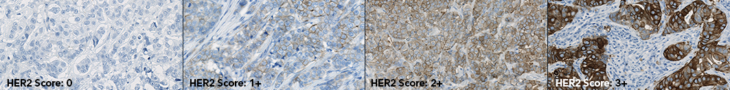
Jan Trøst: Certainly. Breast and gastric cancer tissues possess intrinsic heterogeneity, with gastric cancer exhibiting the greatest degree of heterogeneity, which poses challenges and is the reason for the differences in HER2 scoring algorithms. Misinterpretation or discrepancies in scoring can lead to false-positive or false-negative test results, which can subsequently result in an incorrect treatment decision. It is important to keep this in mind that the reported test result can have significant implications for the patient. Several papers and results from proficiency testing have documented discrepancies in scoring between laboratories, which emphasizes the need for new tools and techniques that can improve accuracy and reproducibility. It is always important to keep in mind that what is done in the laboratory can have a direct impact on patient treatment and outcomes.
“Misinterpretation or discrepancies in scoring can lead to false-positive or false-negative test results, which can subsequently result in an incorrect treatment decision.“
“Several papers and results from proficiency testing have documented discrepancies in scoring between laboratories, which emphasize the need for new tools and techniques that can improve accuracy and reproducibility.“
Visiopharm: Why are low levels of HER2 clinically relevant now?
Jan Trøst: HER2-low is now relevant because a new antibody-drug conjugate, trastuzumab deruxtecan, has demonstrated efficacy in patients with metastatic breast cancer with low HER2 expression, defined as IHC1+ or IHC2+, and negative for HER2 gene amplification. Both the FDA and EMA have approved this indication based on data from the DESTINY-Breast04 trial, where trastuzumab deruxtecan demonstrated benefits over chemotherapy in terms of progression-free survival in patients with otherwise limited treatment options. However, the accurate identification of these patients is challenging. One of the challenges in assessing HER2-low status is that current IHC HER2 assays are not designed to distinguish low levels of HER2 expression. Recent data have shown a 41% discordance among pathologists in distinguishing between IHC0 and IHC1+. This is not sufficient, so there is room for improvement in both assay technology and training, potentially with the support of digital pathology tools.
“One of the challenges in assessing HER2-low status is that current IHC HER2 assays are not designed to distinguish low levels of HER2 expression. Recent data have shown a 41% discordance among pathologists in distinguishing between IHC0 and IHC1+.”

Visiopharm: Can you elaborate more on companion diagnostics?
Jan Trøst: Absolutely. Companion diagnostics belong to the group of predictive biomarkers. While there are various types of biomarkers, such as prognostic and diagnostic biomarkers, predictive biomarkers can predict the outcome related to a specific event, such as a pharmacological intervention. The FDA and EMA define a companion diagnostic as an in vitro diagnostic device that provides information that is essential for the safe and effective use of a corresponding therapeutic product. Companion diagnostic assays has demonstrated their value in both drug development and in the treatment of patients in clinical settings for the past 25 years. These assays play a crucial role, particularly in the case of cancer, where early diagnosis and early intervention are vital, and companion diagnostics help to select the right treatment. In the US, between 60 and 70 drugs or drug combinations have a companion diagnostic assay linked to their use, primarily in oncology and hematology. It is essential that the companion diagnostic assay is developed in parallel with the drug to obtain simultaneous regulatory approval, so it is available in the clinic to support patient selection. However, in recent years, targeted drugs have been approved without a companion diagnostic despite the use of a predictive biomarker assay during clinical development. This discrepancy prompted me to write an article addressing this concern, which was published in the Journal of Clinical Oncology Precision Medicine last year.
“It is essential that the companion diagnostic assay is developed in parallel with the drug to obtain simultaneous regulatory approval, so it is available in the clinic to support patient selection.“
The development of companion diagnostics has primarily been driven by genomic biomarkers, utilizing technologies such as in situ hybridization, polymerase chain reaction, and next-generation sequencing, which account for approximately 75% of the analytical platforms. The remaining 25% is covered by IHC . However, we might see a shift towards protein-based assays in the future. The complexity of the human molecular landscape is substantial, with approximately 22,000 coding genes and with an exponentially growing numbers in the transcriptome and proteome, with approximately 100,000 transcripts and 1,000,000 proteins, respectively. The National Cancer Institute has recently advocated for the integration of proteomics with genomic data, termed “proteogenomics,” to address the additional complexity that proteins and their post-translational modifications add, which are not fully captured by genomic data. Furthermore, proteins are the primary targets of pharmacological interventions.

Visiopharm: What should we expect in terms of future research in this area?
Jan Trøst: When it comes to drug targeting HER2, it looks interesting. A recent article published in Nature Review Drug Discovery discussed HER2 targeted therapy and its future direction and it looks like we should expect a lot. The drug pipelines across various pharmaceutical and biotech companies are brimming with innovations. In the years to come we will likely see the introduction of new monoclonal antibodies, bispecific antibodies, and newer antibody-drug conjugates like trastuzumab deruxtecan and trastuzumab emtansin. Additionally, new tyrosine kinase inhibitors, and therapeutic vaccines are also to be seen in the horizon.
“The drug pipelines across various pharmaceutical and biotech companies are brimming with innovations.”
HER2 targeted drugs are currently used to treat breast, gastric, and esophageal cancer, and recently also non-small cell lung cancer. However, HER2 overexpression is found in several other solid tumors, such as colorectal, bladder, cervical, and biliary tract tumors. Preliminary data from the DESTINT-PanTumor02 trial presented at the recent ASCO meeting showed high response rates, particularly in patients with IHC3+ tumors. Furthermore, a number of other trials with trastuzumab deruxtecan are currently ongoing in patients with different HER2 solid tumors; so, we might expect to see several new indications approved for HER2 targeted therapy in the future.
Visiopharm: How do you see the role of digital tools and AI in diagnostics and treatment?
Jan Trøst: With respect to the utilization of slide-based assays, I foresee a promising future for the integration of digital tools and AI. For IHC and ISH assays, several challenges exist, including issues related to reproducibility and repeatability. I am confident that the implementation of image analysis and AI will help to mitigate these concerns and improve the overall accuracy and reliability of these assays. I think we can say that digital pathology adds a new dimension to this type of assay. For companion diagnostics, incorrect test results can have significant implications on patient safety, and it is imperative to ensure the accuracy and reliability of these assays to avoid potentially harmful treatment errors.
“For IHC and ISH assays, several challenges exist, including issues related to reproducibility and repeatability. I am confident that the implementation of image analysis and AI will help to mitigate these concerns and improve the overall accuracy and reliability of these assays.”
Visiopharm: Thank you Dr. Jan Trøst for sharing your knowledge and experience with us in this insightful conversation.
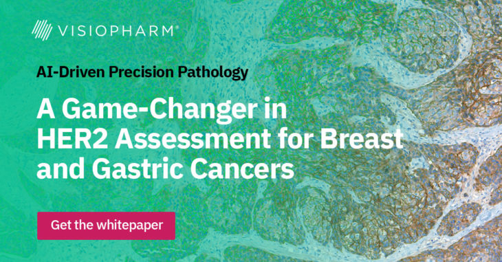
Curious to learn more about Visiopharm’s AI-driven precision pathology solution for HER2?
Contact us here, and download our whitepaper to learn more about Visiopharm’s continuous scoring of the connectivity of the membranous HER2 staining.
Main references related to the interview with Dr. Jan Trøst Jørgensen
- Jørgensen JT, Winther H, Askaa J, Andresen L, Olsen D, Mollerup J. A Companion Diagnostic with Significant Clinical Impact in Treatment of Breast and Gastric Cancer. Front Oncol. 2021; 11: 676939. Read more
- Jørgensen JT. Twenty-five years with HER2 targeted therapy. Ann Transl Med 2023. doi: 10.21037/ atm-23-153. Read more
- Jørgensen JT, Hersom M. Clinical and Regulatory Aspects of Companion Diagnostic Development in Oncology. Clin Pharmacol Ther. 2018;103: 999-1008. Read more
- Jørgensen JT. The potential of trastuzumab deruxtecan as a tissue agnostic drug. Oncology. 2023. doi: 10.1159/000533866. Epub ahead of print. Read more
- Modi S, Jacot W, Yamashita T, Sohn J, Vidal M, Tokunaga E et al.; DESTINY-Breast04 Trial Investigators. Trastuzumab Deruxtecan in Previously Treated HER2-Low Advanced Breast Cancer. N Engl J Med. 2022; 387: 9-20. Read more
- Helwick C. Challenges of Accurately Identifying HER2-Low Breast Cancers. The ASCO Post. February 25, 2023. Read more
- Jørgensen JT. Oncology drug-companion diagnostic combinations. Cancer Treat Res Commun. 2021; 29: 100492. Read more
- FDA. List of Cleared or Approved Companion Diagnostic Devices (In Vitro and Imaging Tools). Read more
- Jørgensen JT. Missing Companion Diagnostic for US Food and Drug Administration-Approved Hematological and Oncological Drugs. JCO Precis Oncol. 2022; 6: e2200100. Read more
- Jørgensen JT. The current landscape of the FDA approved companion diagnostics. Transl Oncol. 2021; 14: 101063. Read more
- Rodriguez H, Zenklusen JC, Staudt LM, Doroshow JH, Lowy DR. The next horizon in precision oncology: Proteogenomics to inform cancer diagnosis and treatment. Cell. 2021; 184: 1661-1670. Read more
- National Cancer Institute. Molecular Diagnostics for Cancer Treatment: Completing the Picture. Read more
- Swain SM, Shastry M, Hamilton E. Targeting HER2-positive breast cancer: advances and future directions. Nat Rev Drug Discov. 2023; 22: 101-126. Read more
- Meric-Bernstam F, Makker V, Oaknin A, Do-Youn O, Banerjee SN, Martin AG, et al. Efficacy and safety of trastuzumab deruxtecan (T-DXd) in patients (pts) with HER2-expressing solid tumors: DESTINY-PanTumor02 (DP-02) interim results. J Clin Oncol. 2023;41 (suppl 17; abstr LBA3000). Read more
 Visiopharm
Categories: Blog
20998
Q&A: HistologiX discusses their journey with Visiopharm’s image analysis solution.
Visiopharm
Categories: Blog
20998
Q&A: HistologiX discusses their journey with Visiopharm’s image analysis solution.
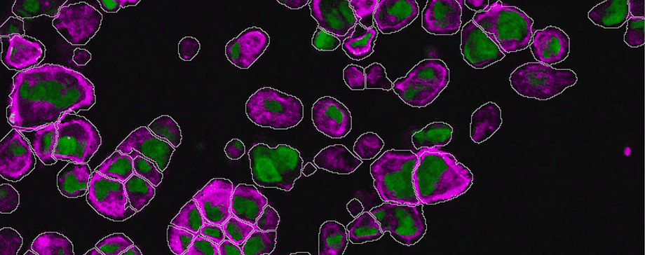
Today, we delve into a conversation with HistologiX, a prominent UK-based CRO that has adopted Visiopharm software to supercharge its image analysis capabilities and add value to its customers. HistologiX specializes in histology and immunohistochemistry services for pharma and biotech. Recently, their digital pathology team has been on a quest for a software solution that enhances their offering and optimizes their capabilities. Let’s learn more about their journey to find the ideal image analysis solution, the hurdles they once faced, and the enhanced offerings they can now present to their customers

James Clay is the Digital Pathology Manager at HistologiX where he developed their digital pathology capabilities, integrating whole slide scanning with quantitative image analysis. He began his scientific career in diagnostic histopathology, specializing in immunohistochemistry and routine histopathology. After a move to contract research, he took lead roles in the design, conduct and reporting of regulatory (GLP) Tissue cross-reactivity studies with novel therapeutic antibodies.

Connor McCracken is a Research Scientist at HistologiX within the Digital Pathology team. Connor’s primary role is in Image Analysis, using AI-driven software to deliver quantitative endpoints for a range of projects, from tissue morphometry to multiplex immunofluorescence. Connor has a 1st Class Bachelors in Biology with a focus on bioinformatics, and is currently undertaking a part-time Masters in Computer Science.
Visiopharm: Welcome, James and Connor. Can you tell us about HistologiX, your services, and how you differentiate yourself from other CROs?
James Clay: We are a UK-based CRO, heavily concentrating on preclinical and developmental studies. From routine toxicology to complex assays like chromogenic and multiplex fluorescence. We do quite a lot of tissue cross-reactivity studies of therapeutic monoclonal and novel antibody constructs like nanobodies for regulatory submissions, and FDA drug submissions. In the last couple of years, we’ve been focusing much more heavily on immunofluorescence and multiplexing. We have a lot of fourplex assays up and running as well as Ultivue’s Immuno8 panel and have recently been doing studies where we are multiplexing RNAScope with IHC immunofluorescence.
Connor McCracken: There’s been a big demand from clients for us to use image analysis on their studies to gain as much information as possible. We are offering it as a service for most studies, especially the ones where the images are quite complex and there’s lots of data to generate.
James Clay: Our distinction lies in our bespoke client-focused approach. Answering the specific questions and challenges the clients have, rather than offering a big bucket list of our services. We get heavily involved with the study design, concepts, data outputs, and questions that they have. It’s always a two-way approach to working with the clients.
Visiopharm: What problems were you trying to solve and what was the limitation you experienced?
Connor McCracken: The main thing we were also missing was flexibility with the platform. With the solutions we had before, the modules are built in a way that “it’s for this or it’s for that”, whereas we were getting questions from clients that required a very specific approach that was not possible with what we had. We couldn’t answer some of the questions we were getting asked from clients unless we had the full open tool set available to us. For instance, in one study, we collected muscle biopsies and had to segment these muscles into individual fibers and then detect RNAscope signals within them, but also a membrane-based protein marker. This tailored analysis was previously impossible because we couldn’t connect these different analyses.
James Clay: We already had analysis capabilities with existing platforms, but one of the major things we lacked was AI/deep learning capabilities, which feed heavily into ROI (region of interest) segmentation, mining the images as much as possible, increasing the reliability of nuclear detections, and things like that. That was one of the main areas we were lacking in our current toolsets. We were always heavily dependent on excessive amounts of manual annotation, which is very time-consuming.
“After trying multiple providers, Visiopharm stood out with its intuitive design and efficient workflows.”
– James Clay, HistologiX
Visiopharm: How did Visiopharm’s software fulfill your needs? Which features were most relevant for you?
Connor McCracken: Having flexibility now with Visiopharm in the way that you can build your own apps, either using one of the prebuilt ones or building it from scratch and then chaining them together in a very nice and logical way – that’s the kind of thing we were missing before. Being able to go under the hood of algorithms is what we wanted and what we’ve got with Visiopharm’s Oncotopix Discovery. We can now just tell our clients “give us your question and we will figure something out, that meets it specifically.” Now we are able to offer something bespoke, that fits whatever questions our clients have.
James Clay: Primarily, just that ability to go beyond the pre-canned algorithms and develop things that were much more biologically relevant to the questions that our customers were asking. Offering individual solutions, which aligns very well with the way we like to work with the clients.
Connor McCracken: Also, the ease of training and applying deep learning has been really great. It’s super easy to use because there’s the pre-trained nuclear segmentation, which works nicely, but it’s easy to go and add extra training regions. Moreover, if we’re building the deep learning from scratch, it’s a similar workflow and we’ve been really impressed with how quick and easy it has been to work with it. We were concerned that it would be hard to work with it and would take lots of training, but it’s been very easy.
James Clay: After trying multiple providers, Visiopharm stood out with its intuitive design and efficient workflows.
Connor McCracken: Additionally, it kind of extends beyond just the software itself, but the support is really good. The training we’ve had from the academy team has been really good and really in-depth. And now, we just go to them with questions, and they just work through it with us and that’s been really helpful. A first-rate service really.
“With Visiopharm’s AI, we can now offer more in-depth and histologically relevant data, more structures, and functional regions of interest.”
– Connor McCracken, HistologiX
Visiopharm: How does Visiopharm’s software impact the way you now work with clients?
Connor McCracken: There are studies where everything, from tissue segmentation to nuclear detection benefits from having AI. Highly accurate nuclear segmentation can really increase the integrity of the data, especially for complex fluorescent samples. With Visiopharm’s AI, we can now offer more in-depth and histologically relevant data, more structures, and functional regions of interest. Moreover, our client’s needs are evolving, they want more complex questions to be answered through images rather than just a very general quantification. Now, we can offer that.
Another study we had was a multiplex IF with a variety of immune and macrophage markers in a xenograft model. We had planned to use Pan-Cytokeratin as a tumor marker, but realized after staining, that 50-75% of the tumor had differentiated to a state, that it was no longer expressing PanCK.Before Visiopharm, this would have meant weeks of manual annotations.
But now, we just used the Tissuealign feature to align the IF and the H&E image. Using deep learning to find the tumor allowed us to exclude other tissue regions like muscle, so we ended up with better information than we would have had just using PanCK. After detection, the regions transferred nicely onto the fluorescent slide, so that we were then able to quantify and phenotype the cells in the tumor/stroma compartments more accurately.
“Before we were always having to fit the study design to the analysis capabilities, whereas now we can fit the analysis approach to the requirements of the study.”
– James Clay, HistologiX
James Clay: This refined methodology has wide applications. We’ve begun exploring its use in multiplex fluorescence. By integrating registered H&E in the same section or adding other fluorophores, we can provide more cellular histological context. This level of detail, especially when combined with deep learning, enables more refined regions-of-interest analysis and a higher data granularity. We are doing many more studies now in the spatial biology sphere, like immune-oncology with lots of multiplex markers. The ability to build the spatial relationships how we want, not how it is pre-canned in an algorithm already, is really helpful. Visiopharm has transformed our approach. Not only does it enable deeper morphometric analysis, but it also allows for more precise quantification of protein expression within detected objects. One of the driving factors for our switch was the software’s ability to offer in-depth customized filtering, something our previous solution didn’t provide. But mostly, being able to design the right tool for the right job.
Before we were always having to fit the study design to the analysis capabilities, whereas now we can fit the analysis approach to the requirements of the study.
Transform your image analysis approach with Visiopharm
Navigating the complex waters of histology and immunohistochemistry requires more than just knowledge—it demands the right tools. In a rapidly evolving market landscape, a flexible software solution is not just an asset—it’s a competitive edge. The transformative journey of HistologiX stands as a blueprint for the potential of the right technological integration. The adoption of Visiopharm has been more than a mere upgrade—it’s been a catalyst.
The benefits for the CRO have been manifold:
- Precision & Efficiency: From reducing manual annotations to implementing AI for tissue classification, the software has significantly streamlined workflows, allowing the CRO to deliver results with greater accuracy in reduced timeframes.
- Client-centric Solutions: With the flexibility of Visiopharm, the CRO can now tailor solutions to address specific challenges posed by clients. Whether it’s building from scratch or chaining algorithms, the CRO has newfound agility to meet diverse analytical needs.
- Business Growth & Diversification: The CRO’s capability to tackle more complex questions and provide detailed image analyses, facilitated by Visiopharm, opens doors to new project types and a broader client base.
If you would like to learn more about the flexibility of Visiopharm software, contact us here.
Curious to learn more about HistologiX’s services in histology and IHC? Get in touch with the HistologiX team at info@histologix.com or visit their website for more information.
 Visiopharm
Categories: Blog, Customer Stories
21052
NPIC and Visiopharm collaborate to advance H&E staining standardisation
Visiopharm
Categories: Blog, Customer Stories
21052
NPIC and Visiopharm collaborate to advance H&E staining standardisation
The National Pathology Imaging Co-operative (NPIC) of Leeds Teaching Hospitals NHS Trust, and Visiopharm, a leading provider of AI-driven precision pathology software, jointly announce a collaboration dedicated to supporting the standardisation of Haematoxylin and Eosin (H&E) staining.
Harnessing their combined expertise, NPIC and Visiopharm are developing an innovative solution that brings together ‘Tango’, NPIC’s tissue-mimicking stain assessment slide, and a fully automated image analysis algorithm developed by Visiopharm. The algorithm will be made available within Visiopharm’s Qualitopix™ platform as part of NPIC’s imminent UK National Stain Survey, where labs can quantitatively measure H&E staining intensity over time.
Staining tissue in histopathology is an important step to allow visualisation of cellular structures, but staining can be variable, and while humans are able to tolerate a wide level of variation, computer analysis can be thrown by this variation. Computational methods can be used to improve AI performance, including colour normalisation, but ultimately these are arbitrary. NPIC’s Tango tool provides an absolute reference, enabling the creation of more consistent data for AI development, with the aim of improving patient care and speed of cancer diagnosis in the future. Not only can Tango help improve AI, but it can also improve stain utilisation in the laboratory, potentially saving costs due to reduced reagent waste.
Dirk Vossen, Chief Diagnostics Officer at Visiopharm, shares: “This collaboration with NPIC signifies an important step towards addressing the root causes of H&E staining variations, which can influence both human interpretation and AI-based assessments in histopathology. Our combined solution, integrating Tango and automated analysis on our Qualitopix platform, offers a much-needed standardisation enabler in the field.”
David Brettle, Chief Scientific Officer at Leeds Teaching Hospitals NHS Trust and NPIC’s Quality Co-ordination Centre lead, adds: “We are delighted to be collaborating with Visiopharm taking Tango, our on-slide H&E stain assessment tool, from the research lab into a viable real-world platform. This work will allow optimisation and standardisation in the laboratory and enable the potential for guidelines and standards to be produced, improving not only the quality of slides, but also the digital image and ultimately supporting effective and efficient AI development and deployment.”
It is anticipated that following validation, the analysis algorithm will become accessible to all Visiopharm customers on the Qualitopix platform, extending its utility and impact within the histopathological community, ready for when the Tango stain assessment slides become commercially available.
About NPIC
The National Pathology Imaging Co-operative (NPIC) is supported by a £50m investment from the Data to Early Diagnosis and Precision Medicine strand of the government’s Industrial Strategy Challenge Fund, managed and delivered by UK Research and Innovation (UKRI). NPIC is a unique collaboration between NHS, Academic and Industry partners with aims to: deploy digital pathology systems in hospitals across the country to improve pathology services and patient outcomes, disseminate best practice in digital pathology, develop and evaluate AI in pathology, and create a platform for nationwide research and innovation.
NPIC’s Quality Co-ordination Centre (QCC) aims to understand and quantify the variations in digital pathology images, enhance international quality standards and provide resources to encourage high tissue-to-image fidelity and production of high-quality digital pathology image datasets. NPIC’s QCC also want to encourage good practice in the digital pathology community and to drive the quality improvement cycle in digital pathology.
About Visiopharm
Visiopharm® is a leading provider of AI-driven precision pathology software for research and diagnostics. In research, it is a technology leader providing tools that help scientists, pathologists, and image analysis experts produce accurate data for all types of tissue-based research. In diagnostics, it is a leader within clinical applications, with no less than eight diagnostic algorithms cleared under IVDR for EU and UK customers. These applications provide diagnostic decision support and and can be easily activated and integrated into existing lab workflows.
Founded in 2002, Visiopharm is privately owned and operates internationally with over 750 customer accounts in more than 40 countries. The company’s headquarters are located in Denmark’s Medicon Valley, with offices in Sweden, the UK, Germany, the Netherlands, and the United States, and local representation in France and China.
Contact:
Visiopharm:
Johanne Louise Brændgaard
Chief Marketing Officer
jlb@visiopharm.com
NPIC:
Catherine Colquhoun
Innovation Project Manager
leedsth-tr.npicresearch@nhs.net
 Visiopharm
Categories: Press Releases Tags: Qualitopix H&E
Visiopharm
Categories: Press Releases Tags: Qualitopix H&E