Stay curious

As imaging grows more complex, fast and precise computational analysis is essential.
Yet, one-size-fits-all tools fall short, and piecing together DIY pipelines is slow and expertise‑heavy. You don’t want to analyze images; you want reliable data that moves your research forward.
Our flexible image analysis software lets you build tailored analyses to generate the data you need. For drug discovery and development, spatial biology, or clinical research.
Turning images into knowledge
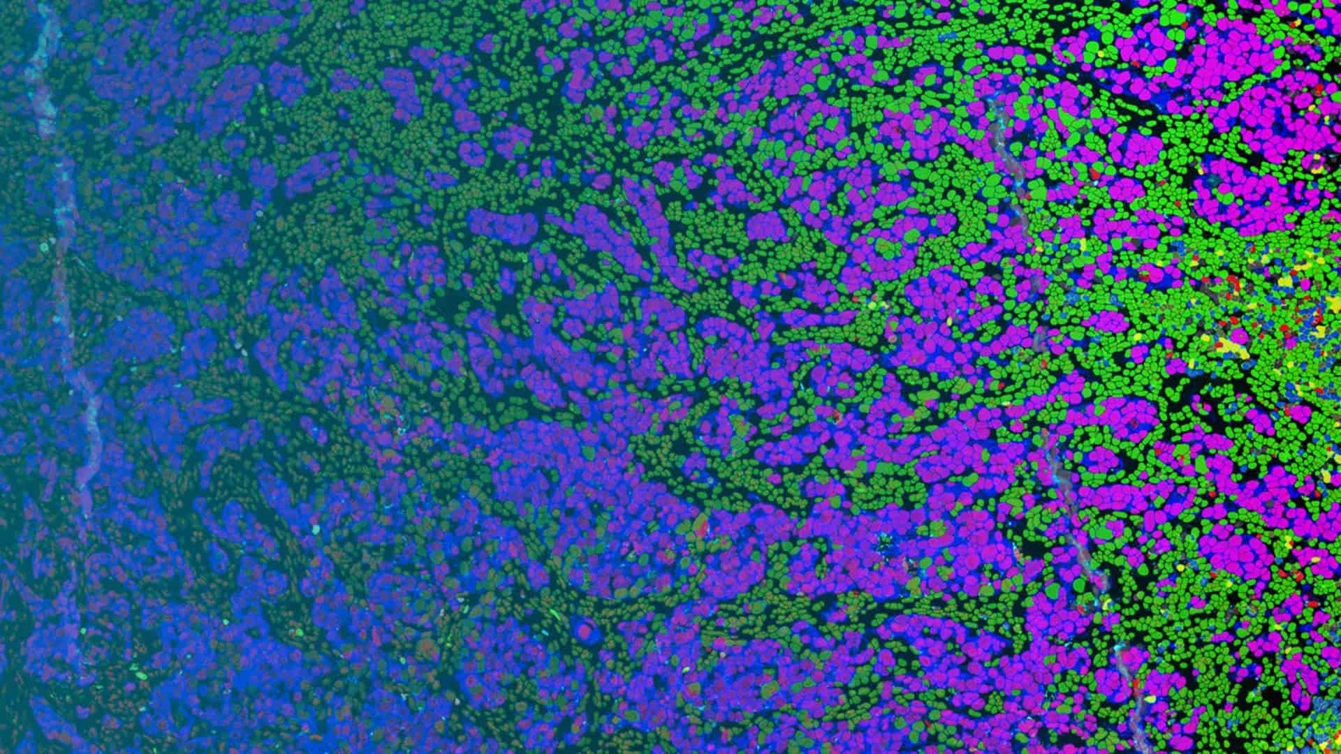
Spatial Biology Analysis
A complete workflow for all your
multiplexed image analysis needs.
One platform. Multiple modalities. More answers.
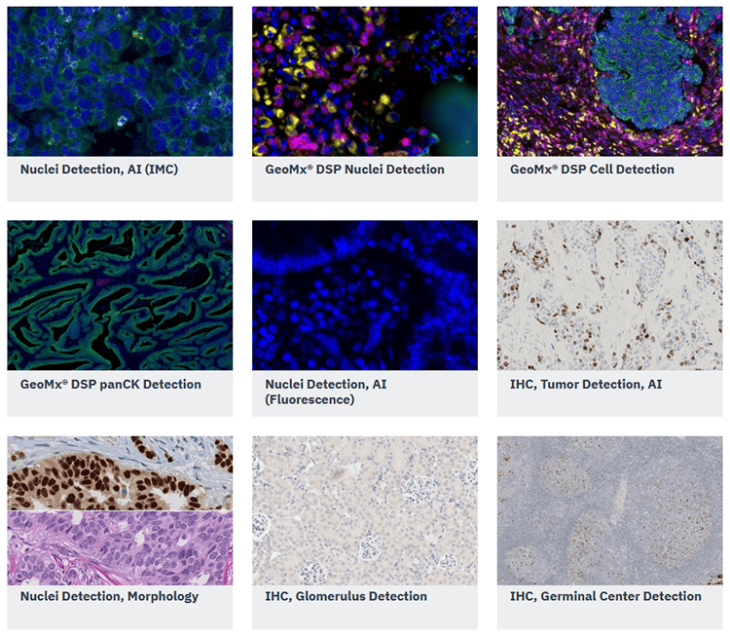
Tissue Image Analysis
Turning expert knowledge into reliable results through intuitive image analysis.
.

“This highlighted one of the main benefits of using Visiopharm: which is the flexibility we have with APP design.”
Karen McClymont, Image Analysis Project Manager at OracleBio

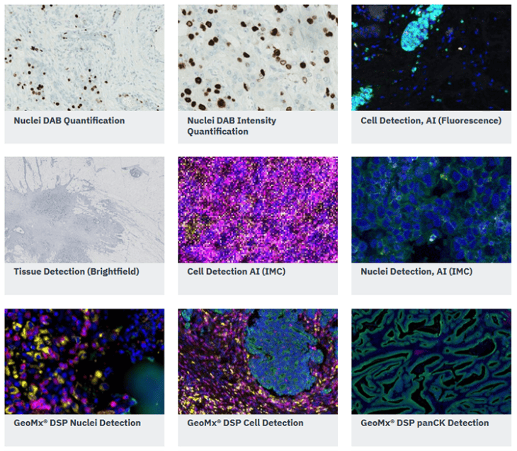
Get started easily with our Quickstart APPs
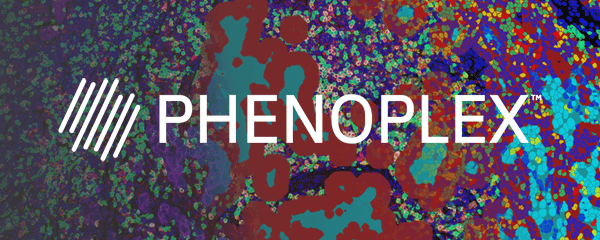
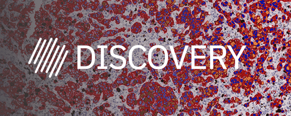
Image analysis doesn’t have to slow you down. Discovery and Phenoplex include Quickstart APPs, our preconfigured tools for segmentation tasks from tissue detection to various region detections and to cell and nuclei detections. Ready in just a few clicks across different imaging modalities.
Start immediately, and refine results further with supervised deep learning for maximum accuracy.
Customizable
Mix and match to create your own workflows. Adjustable with evolving needs.
Versatile
Supports datasets from multiple imaging modalities.
Built into your plan
Included in your Discovery and Phenoplex software subscription.
Real world applications of our software
Our image analysis software supports scientists across all indications.
Explore how they are using Visiopharm, from basic research to developing diagnostic LDTs.
Click the dots to learn more
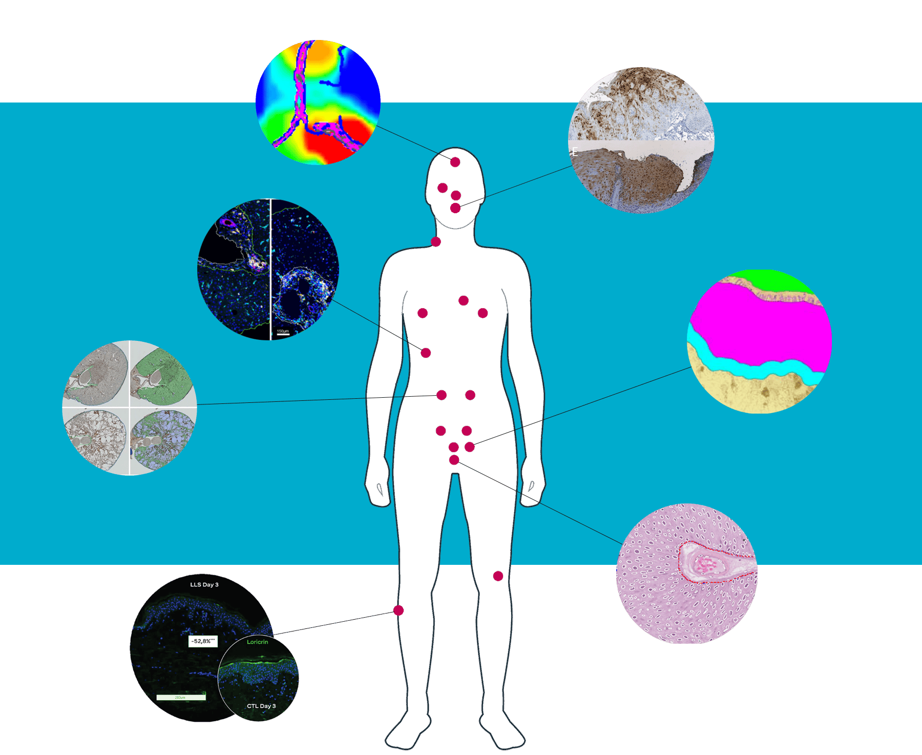
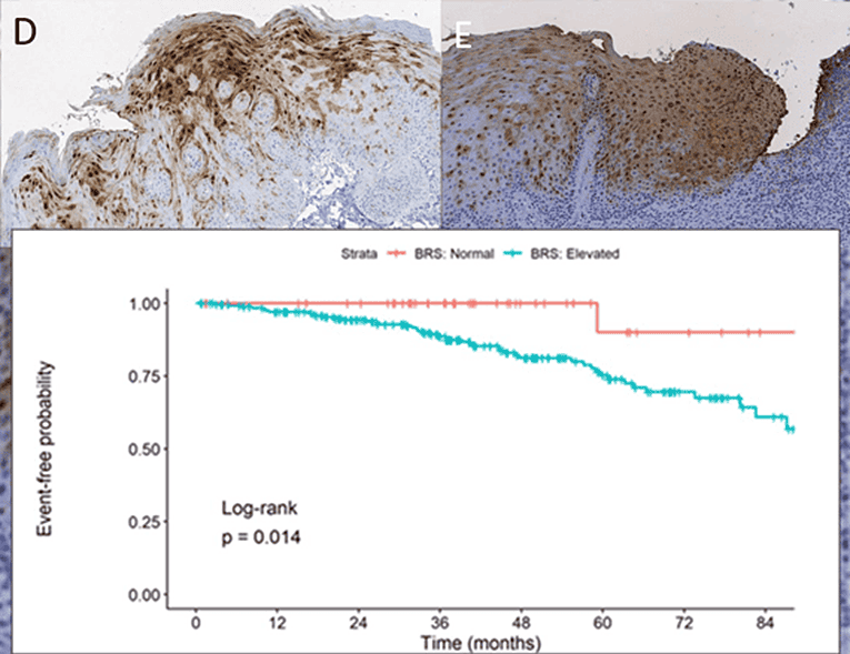
Oral cancer laboratory developed test (LDT)
Oral cancer
Proteocyte developed an APP with the Discovery platform to predict the development of high risk lesions. Validated as an LDT with 96.2% sensitivity and negative predictive value.
Detecting Parkinson’s 15 years in advance
Neuroscience
CND Life Sciences turned biomarker research into risk identification using the Discovery platform. They created an APP that measures the occurrence of the highly elusive biomarker P-SYN. It is now widely used by the company’s pathologists.
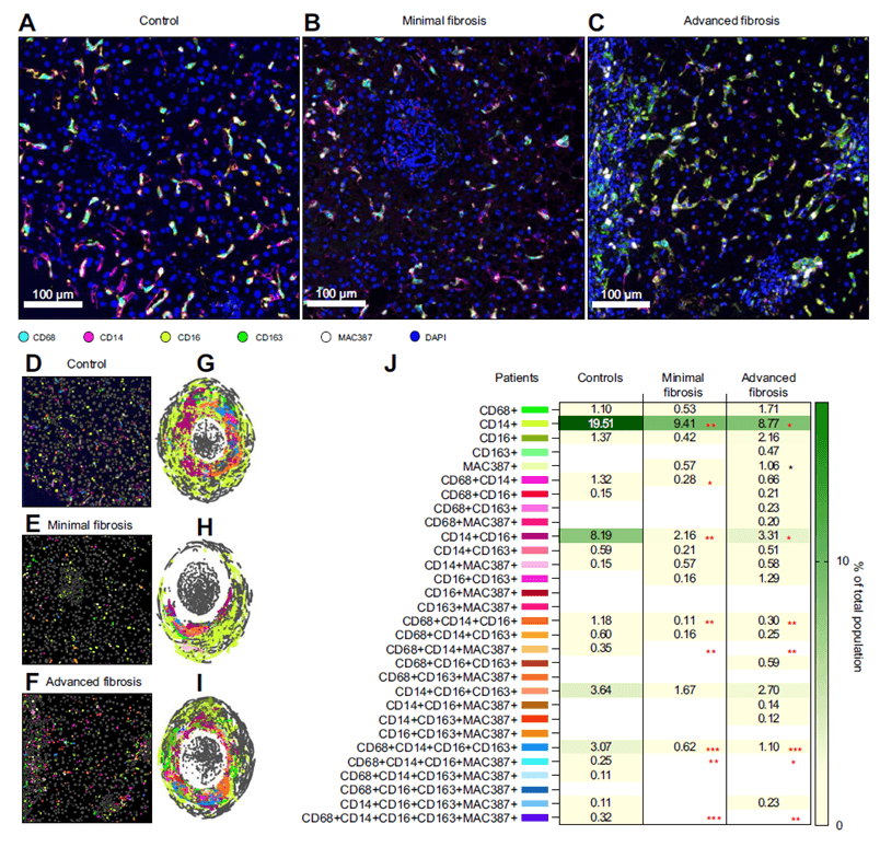
Macrophage diversity in liver disease
Hepatology
At the University of Texas Medical Branch, Dr. Heather Stevenson-Lerner uses Visiopharm’s Phenoplex to quantify immune cells in fibrotic liver diseases. The spatial analysis reveals complex macrophage patterns—advancing patient stratification and precision medicine.
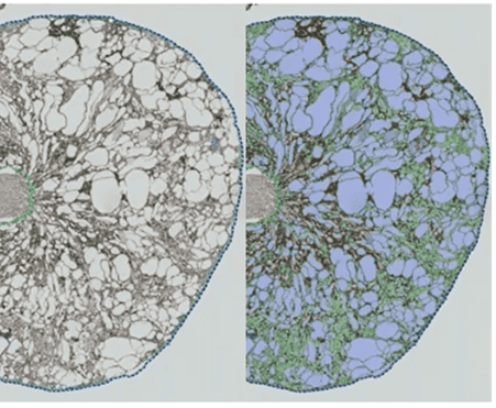
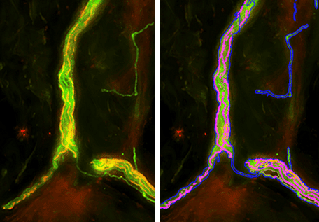
Quantification of kidney cysts
Nephrology
Novalix uses a PKD1-inducible knockout mouse and Visiopharm’s Discovery to precisely quantify cysts, vessels, and fibrosis over time. This enables robust tracking of disease progression and therapeutic response.
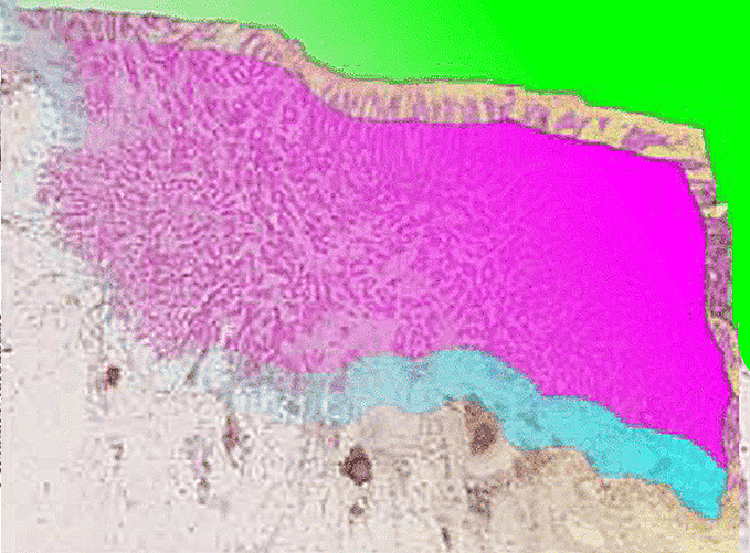
Lymphocyte infiltration in colorectal cancer
Colorectal cancer
Anne-Marie K. Fiehn’s team in Copenhagen used Visiopharm’s deep learning APPs to segment colorectal cancer tissue into core and margin regions, enabling automated quantification of invasive lymphocytes.
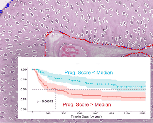
Reducing inter-observer variability in cancer grading
Bladder cancer
Even the most experienced pathologists come to different conclusions in grading bladder cancer up to 20% of the time. The Berman Lab wanted to fix that by training an algorithm on objective features within nuclei. The result was a clearer distinction between high- and low-grade cancer.
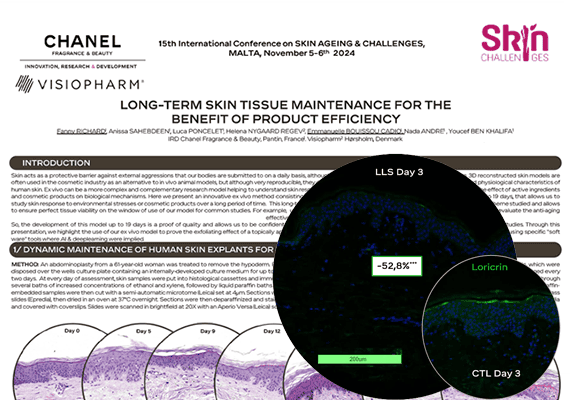
The proof is in the protein
Dermatology
Using the Discovery platform, Chanel measured the efficacy of a novel, exfoliating skin product by visualizing 3 proteins associated with exfoliation in an animal-free skin model.
Main benefits of our image analysis software
Flexible analysis tailored to your needs
Our image analysis software is highly flexible, allowing researchers to build custom workflows tailored to their specific questions instead of relying on fixed modules.
Users can start with combining pre‑trained Quickstart Apps for standard tasks. For further refinement retrain on your own images, or extend with additional processing steps or custom export values, resulting in truly bespoke image analysis for any research need.
Trust your results
Any selected data point in your data exploration graphs and plots will link back to the respective cell in the image/TMA to allow quick verification of the results. All plots can be adjusted to display each tissue compartment, phenotype, biomarker, or cell, as needed to properly interrogate results.
See it – train it – find it
With integrated the deep learning, researchers can train the software to detect any region of interest and focus analysis exactly where it matters. Apply your tissue expertise and annotate the regions and structures you want to identify. Our intuitive drawing tool allows for quick and seamless annotation. Leave the rest to Visiopharm.
No coding, no time-consuming training, no image analysis experience required.
Analyze consecutive slides
Align and subsequently analyze serial sections with our Tissuealign™ analysis module. This allows co-localization studies of multiple biomarkers also across image modalities.
Align and co-analyze multiplex IF with H&E or IHC brightfield to get the most out of your data.
Tissue microarrays (TMA)
Tissuearray™ is one of the most comprehensive, dedicated solution available on the market. A guided workflow allows for managing cores and importing grid designs as well as apply any analysis solution in batch processing.
Speed that meets your needs
Run batch processing across entire studies. Our image analysis software makes optimal use of the computational resources available where it is installed, for parallelized, fast processing.