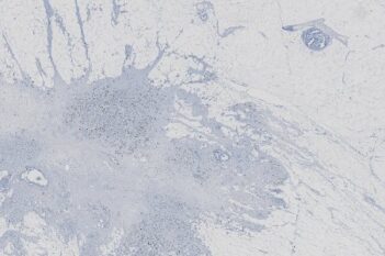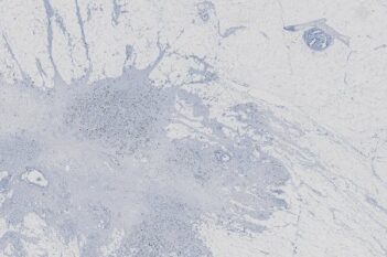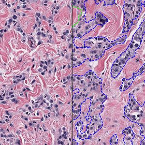
Analysis on breast cancer tissue

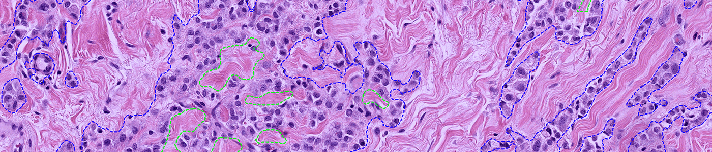
#10197

The H&E Tumor Detection APP uses deep learning to automatically identify and outline tumor regions in H&E-stained brightfield images of different tissue types. It delivers accurate tumor ROIs and a quantitative total tumor area measurement (mm²).
Using our Tissuealign module, users can register the H&E with different stained sections (other brightfield or any IF) to transfer the tumor regions for further detailed analysis onto the aligned stains. This enables downstream analysis being limited to tumor-specific regions, also on stains where tumor detection is not possible by itself.
Trained on a diverse set of tissues and laboratories, the model is robust to staining variability across multiple organs. Users can easily adjust the minimum area of detected tumor or fine-tune the model to their own data, making the app adaptable to a wide range of research applications.
Analysis on breast cancer tissue
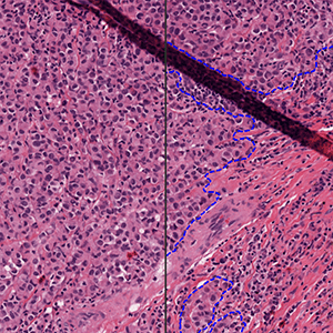
Analysis on melanoma tissue with exclusion of an artefact
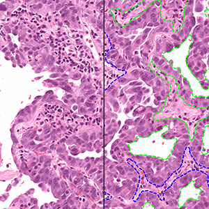
Analysis on lung cancer tissue
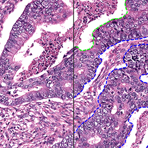
Analysis on colon cancer tissue
Quantitative Output variables
Output is the total tumor area in mm².