
From manual counting to AI-supported assessments: How Växjö Pathology Lab integrated AI into clinical practice
In the conversation below Dr. Kitti Brinyiczki reflects on the team’s experience and the lessons they have learned along the way.
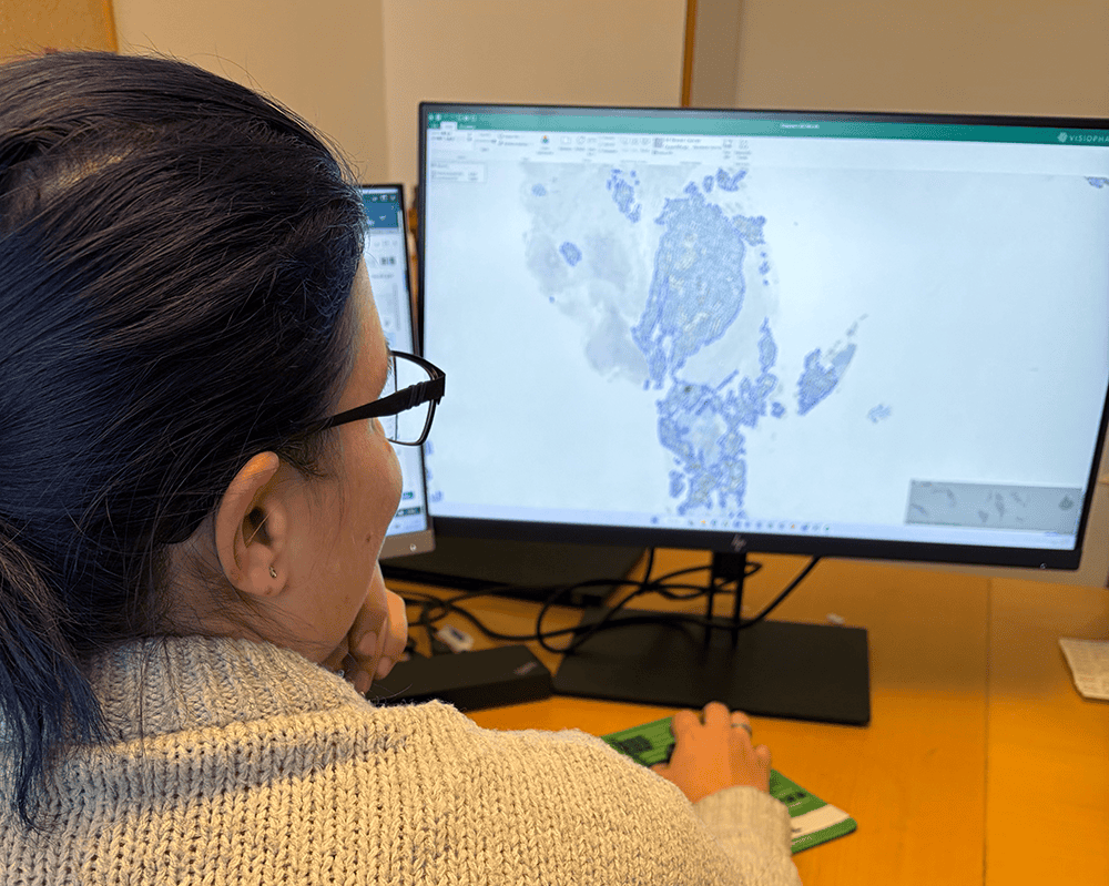
Dr. Kitti Brinyiczki, pathologist from Växjö Pathology Lab.
Tell us about your laboratory and what you work with.
So, the Pathology Department at Växjö Hospital is a Swedish regional pathology lab handling approximately 15,000–16,000 diagnostic cases each year. The department works broadly across general surgical pathology, with particularly high volumes in skin, breast, prostate and gastrointestinal pathology. More complex cases are generally centralized to the major university hospitals. Our team consists of eight pathologists and two pathology residents, some of whom also work with cytopathology. As is common in smaller regional hospitals, pathologists work across several areas rather than being too subspecialized. We have been working with Visiopharm for quite some time and have supported the development of the Ki-67 assessment APP.
What prompted you to explore AI-supported assessment?
Our initial goal was to reduce variation between pathologists’ assessments and make the results more objective. That was what first sparked our interest in using AI. Then, in 2022, the KVAST working group of the Swedish Society of Pathology recommended moving away from counting Ki-67 in hotspots. Instead, we were advised to assess Ki-67 across the entire stained tumour area, a whole-slide Ki-67 assessment, using what is known as a weighted global score, which was first recommended by the International Ki67 in Breast Cancer Working Group in 2021.1
This is still quite difficult to do manually because our eyes are accustomed to searching for hotspots. When we began working with Ki-67 in this way, I became more involved with Visiopharm and had the opportunity to work more closely with the team.
Honestly, it was a really good experience working with you.
How was working with the Visiopharm team, how did you collaborate?
Honestly, it was a really good experience working with you. Whenever I had questions or concerns, I always got help. I genuinely enjoyed the collaboration. During the testing process, I also really had fun, experimenting a lot and trying many different things in the test environment. Communication worked very well and I always received support when I had questions.
What did you learn when you compared AI with manual Ki-67 assessment?
I was not involved in developing the solution itself, but I participated in the testing once it became available. We spent a lot of time testing different slides and staining variations to ensure that the solution worked.
I also conducted a small study here in the laboratory in which I counted Ki-67 in different areas of the slide manually. Even when I deliberately tried to select different areas, I continued to focus on hotspots. I was able to demonstrate this clearly, both to myself and to my colleagues.
What was fantastic was that the Visiopharm APP included a heatmap. It allowed us to compare different areas and see that I still ended up close to a hotspot, even when I tried to select other regions. This demonstrated how challenging it is to assess the entire tumour area manually in the intended way. Our eyes are naturally drawn to areas with high positivity. This makes it difficult to perform a completely objective assessment, and we risk overestimating Ki-67. In some cases, we found that the manual assessment was 5–10 percentage points higher.

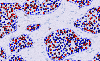
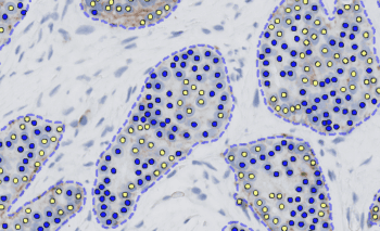
Ki-67 stained breast cancer tissue with classification results of our Ki-67 APP
What would the clinical impact of this difference be?
Ki-67 assessment indicates whether tumor proliferation is low, intermediate or high. When proliferation is low, it may not matter very much whether the result is 1%, 3% or 4%. But it becomes particularly important in cases with intermediate proliferation, where a difference of few percentage points can have a significant impact. Oncologists want to know the proliferation level because it may indicate how the tumor could respond to treatment. Their clinical guidelines describe how patients should be treated according to their Ki-67 and ER/PR results. The assessment therefore matters because it may place a patient in a different treatment group. If the score is 5-10% too high, the treatment group may not be the optimal one.
With AI, I no longer need to sit and count 200 cells manually, which saves me a great deal of time.
How has AI changed your Ki-67 workflow?
It is faster to report the cases, although the time saved varies because pathologists work in different ways. Some estimate the percentage visually, whereas I personally tend to count quite a lot, using a manual counter, because I want to feel confident in my assessment. With AI, I no longer need to sit and count 200 cells manually, which saves me a great deal of time. At multidisciplinary breast meetings, pathologists, surgeons, oncologists and radiologists discuss each patient together. Questions were sometimes raised about the Ki-67 assessment, for example, whether the value should have been higher or lower. Since we began using AI, we feel that these types of questions have become less common.
What role does AI play in your assessments?
Manual assessment is subjective. I may be more tired in the afternoon or on Friday, but AI does not get tired. AI applies the same criteria consistently, which can help reduce differences between observers. When a result leaves the laboratory, it should be as objective as possible. AI is therefore a tool for supporting our assessments. It does not replace pathologists but should serve as an aid.
Experience within breast pathology naturally varies across the team. AI gives everyone access to the same objective support, which can contribute to more consistent results throughout the laboratory. AI acts as a complement to our colleagues’ expertise, particularly in borderline cases. When I am uncertain or close to a cutoff, I may otherwise need to ask a colleague, “Do you also think this is 11% or 20%?” It can be a difficult assessment for us as pathologists, while the result may have considerable significance for the patient and the oncologist.
This is particularly noticeable in HER2 assessments, where we can sometimes feel uncertain. We have three doctors in the laboratory with a strong interest and extensive experience in breast pathology, and we usually turn to them when we have questions. When they are not available, the HER2 APP provides additional support. This has reduced the need to consult them and saves time for both us and even more so for them.
Why is accurate HER2 assessment so important?
HER2 is assessed using immunohistochemical membrane staining and has a direct impact on treatment because HER2-targeted therapies are available. HER2 expression is traditionally scored from 0 to 3+. If a tumour shows very strong positivity and is scored as 3+, the patient may be eligible for treatment directly. Cases scored as 2+ require additional testing to determine whether the HER2 gene is amplified. If amplification is confirmed, the patient may be also eligible for HER2-targeted treatment.
And now different treatments have been developed for patients with so-called HER2-low or HER2-ultralow breast cancer, with specific criteria for these groups. HER2 assessment therefore has a direct and increasing influence on treatment options. The distinction between HER2 1+ and 2+ is particularly important. A 2+ case must be sent for additional testing, whereas a 1+ case is not sent. This is one of the key borderline areas in HER2 assessment.
How did the laboratory’s digitalisation journey prepare you for AI?
In Växjö, we can work entirely digitally. Some of our colleagues work from home several days a week, and we also submit requests for additional tests digitally. The workflow is fully digitalised, which is quite advanced. We began digitalising because we had relatively few doctors and needed to find different ways of working. By working digitally, pathologists in other locations could help with diagnosis. We initially scanned specific cases, but gradually began scanning more and more. Thus, we are very positive towards digitalisation and no one in the team was opposed to AI because everyone understood that it was a support tool that we could choose to use when needed.
We now have a fully automated workflow and do not need to send cases for analysis ourselves. When I open a case, everything has already been counted. It really works as promised and as shown in the videos, the analysis is already complete.
What helped you integrate AI into the routine workflow?
You need someone who is good with IT. We were fortunate to have Ingela Wilkens and Peter Lindblad. They could examine the data and the HTML logs and understand what they meant. Thus, they became an excellent internal bridge between pathologists and IT. We now have a fully automated workflow and do not need to send cases for analysis ourselves. When I open a case, everything has already been counted. It really works as promised and as shown in the videos, the analysis is already complete.
What is important for pathologists to understand when they begin using AI?
AI has limitations. Cases that are difficult for us as pathologists will also be difficult for AI. AI should not replace our diagnostic work but strengthen our assessments. We should not expect AI to do exactly the same thing that I do in my head. AI works in a more mathematical way. After all, we are doctors, not mathematicians, so it can initially be difficult to understand and feel confident in how the calculation works across the entire tumour area. What matters, however, is the proportion of positive and negative cells rather than the exact number of cells counted. If the software analyses three-quarters of the tumour and the cell distribution is representative, it can produce approximately the same percentage as an analysis of the entire tumour and is much more representative than any manual evaluation. When the calculation already includes around 200,000 tumour cells, adding another 2,000 cells will usually have only a limited effect on the final percentage. It can take time for pathologists to become comfortable with this numerical approach to whole-slide assessment. It is simply a different way of thinking.
To summarise, what role can AI play in a pathologist’s daily work?
As humans, we can only make a certain number of decisions before our brains become tired. When we have many cases, we constantly need to make different assessments: What type of tumour is this? Is this a tumour cell or a stromal cell? How thick is the tumour? What is the distance between the tumour and the resection margin? At the same time, we also make ordinary decisions throughout the day, such as what to have for lunch. From the moment we wake up, our brains are constantly working, and eventually they become tired. AI can help by reducing the number of decisions we need to make. Those are decisions that no longer have to occupy our attention, leaving more energy for the truly important diagnostic questions. This leaves us with fewer routine decisions to make and allows us to devote more of our capacity to the more demanding diagnostic assessments.
- Nielsen et al., Assessment of Ki67 in Breast Cancer: Updated Recommendations From the International Ki67 in Breast Cancer Working Group, JNCI: Journal of the National Cancer Institute, Volume 113, Issue 7, July 2021, Pages 808–819, Link
Share this article