Stay ahead

Realize the
Our promise
Built to help you stay ahead
Our unique software combines 20 years of knowledge gained by working with pathologists and research scientists with the power of AI. We provide you with the accuracy and versatility you need to meet the tissue analysis challenges of tomorrow in research and diagnostics. Whether you’re developing next-generation therapies or matching these therapies with the right patients, we ensure the quality you need to stay ahead.
What’s your greatest discovery?
We enable scientists to conduct tailored research with flexible image analysis software

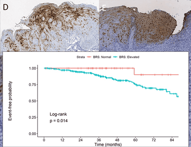
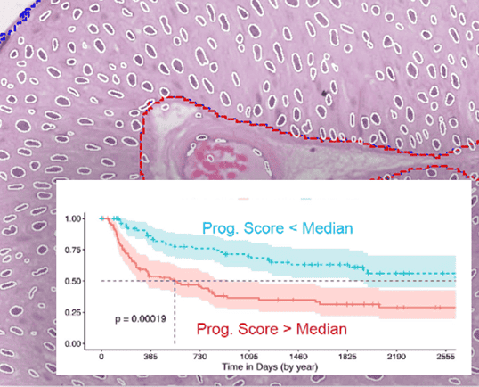
Oral cancer laboratory developed test (LDT)
Oral cancer
Proteocyte developed an APP with the Discovery platform to predict the development of high risk lesions. Validated as an LDT with 96.2% sensitivity and negative predictive value.
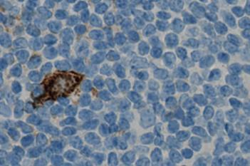
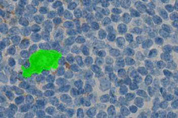
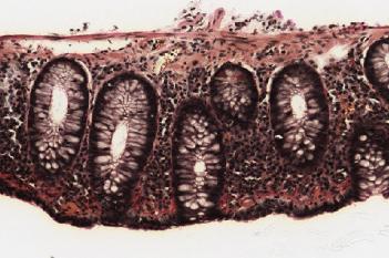
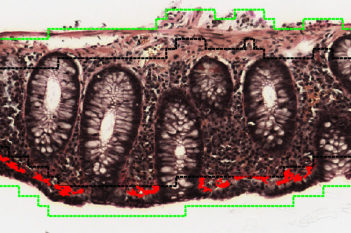
Lymphocyte infiltration in colorectal cancer
Colorectal cancer
Anne-Marie K. Fiehn’s team in Copenhagen used Visiopharm’s deep learning APPs to segment colorectal cancer tissue into core and margin regions, enabling automated quantification of invasive lymphocytes.
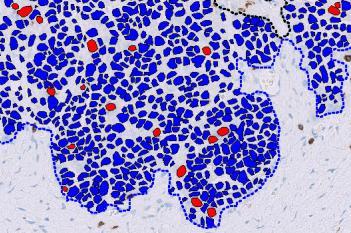
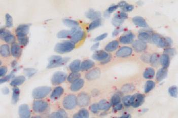
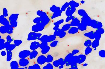
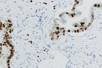
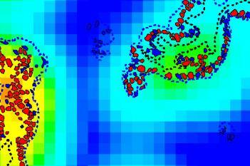
Reducing inter-observer variability in cancer grading
Bladder cancer
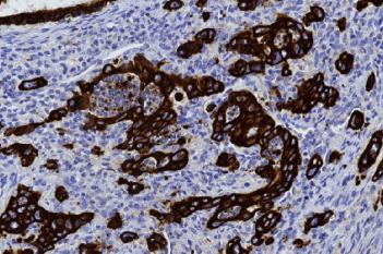
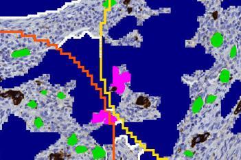
Even the most experienced pathologists come to different conclusions in grading bladder cancer up to 20% of the time. The Berman Lab wanted to fix that by training an algorithm on objective features within nuclei. The result was a clearer distinction between high- and low-grade cancer.
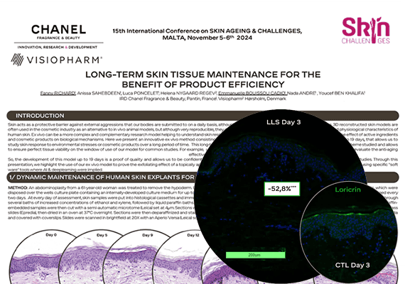
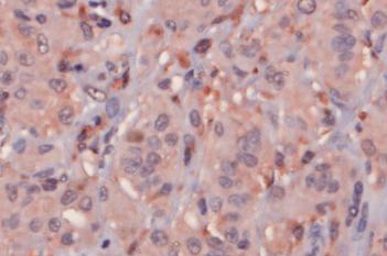
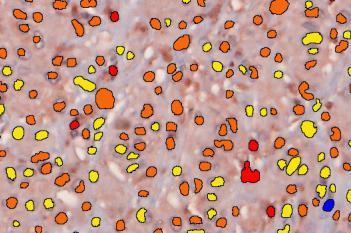
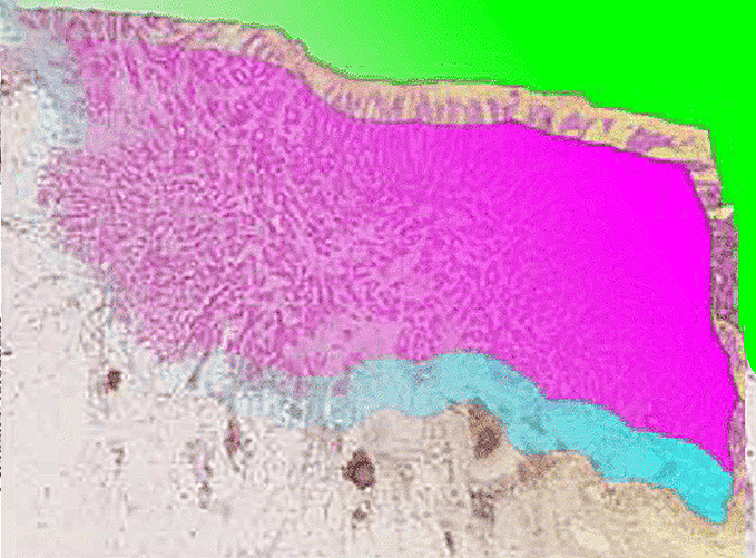
The proof is in the protein
Dermatology
Using the Discovery platform, Chanel measured the efficacy of a novel, exfoliating skin product by visualizing 3 proteins associated with exfoliation in an animal-free skin model.
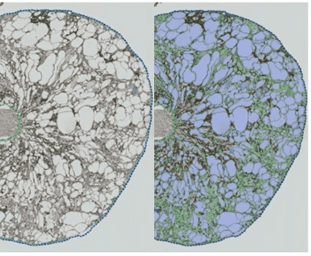
Quantification of kidney cysts
Nephrology
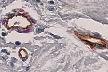
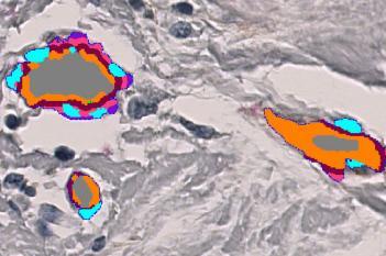
Novalix uses a PKD1-inducible knockout mouse and Visiopharm’s Discovery to precisely quantify cysts, vessels, and fibrosis over time. This enables robust tracking of disease progression and therapeutic response.
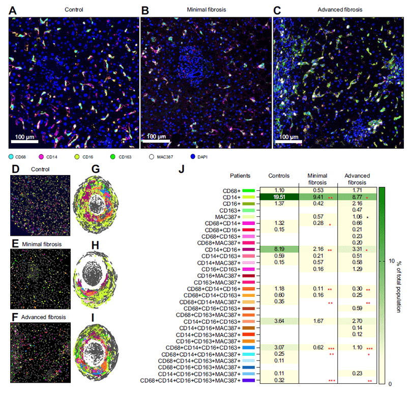
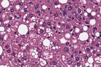
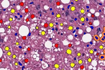
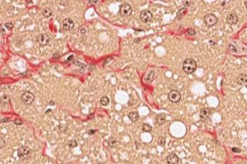
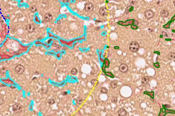
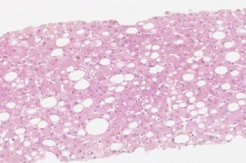
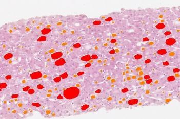
Macrophage diversity in liver disease
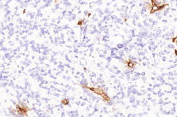
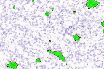
Hepatology
At the University of Texas Medical Branch, Dr. Heather Stevenson-Lerner uses Visiopharm’s Phenoplex to quantify immune cells in fibrotic liver diseases. The spatial analysis reveals complex macrophage patterns—advancing patient stratification and precision medicine.
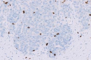
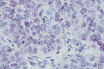
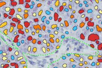
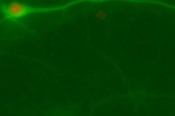
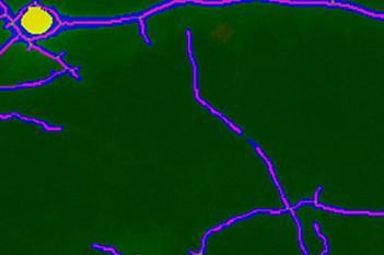
Detecting Parkinson’s 15 years in advance
Neuroscience
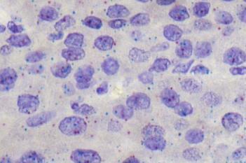
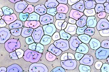
CND Life Sciences turned biomarker research into risk identification using the Discovery platform. They created an APP that measures the occurrence of the highly elusive biomarker P-SYN. It is now widely used by the company’s pathologists.


News

Spatial Biology Analysis
Phenoplex™ is our complete image analysis workflow for all your multiplexed image analysis needs in spatial biology.
Diagnostic decision support
Discover our IVDR certified CE-IVD APPs and our integration workflows that revolutionize standardization through AI-driven pathology solutions.
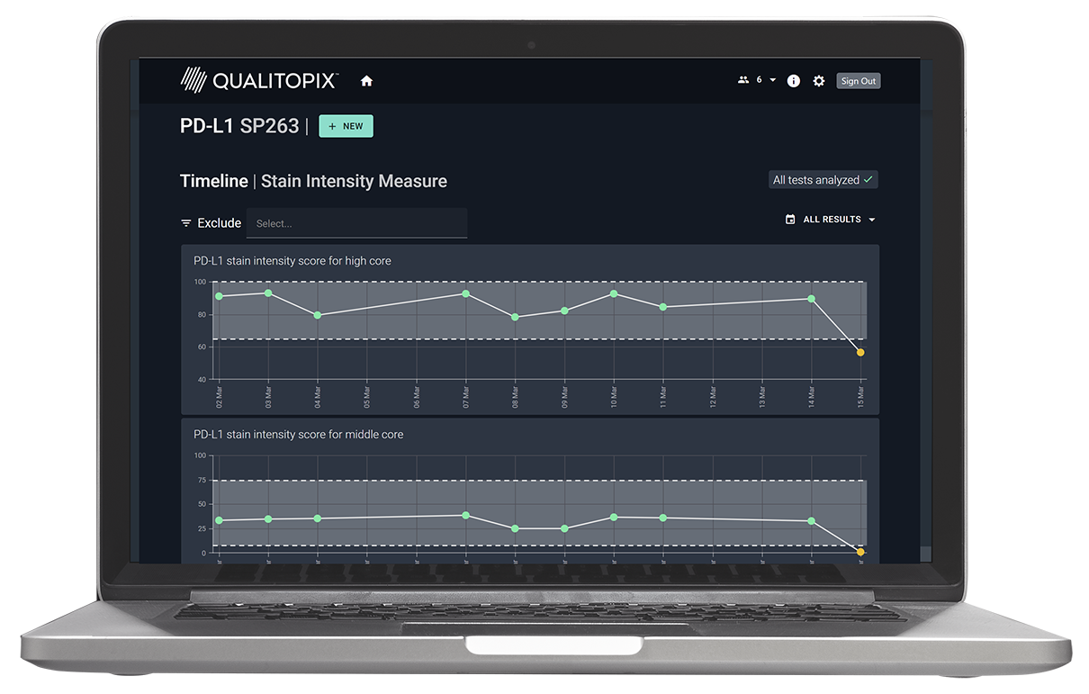
IHC staining consistency
Measure and document your staining consistency through standardized analysis of next generation reference materials with Qualitopix.
Tissue image analysis software
Turning expert knowledge into reliable results through intuitive image analysis.
With Discovery, anyone with an understanding of tissue morphology can train an AI deep learning APP to get accurate and reproducible results.