Q&A: NovAliX’s journey with Visiopharm’s image analysis software
Welcome to our latest blog post, where we delve into the evolving landscape of histology and image analysis through an exclusive interview with industry experts from NovAliX – a fully integrated drug discovery CRO based in Strasbourg, France.
In this insightful conversation, Didier Merciris and Florence Anquetil-Besnard from NovAliX share their experiences in incorporating Visiopharm’s image analysis software into NovAliX’s workflows, and the profound impact it has had on their projects and clients.
Inflammation (Osteoarthritis/Arthritis (joints), Osteoporosis (bone), IBD (colon), Lupus (kidney) and fibrosis (liver, kidney, lung).
Visiopharm: Welcome, Didier and Florence. Can you tell us about NovAliX, the services you offer, and how you differentiate yourself from other CROs?
Florence Anquetil-Besnard: NovAliX is a fully integrated drug discovery CRO. We support biopharma companies with a range of programs, from initial target identification to the delivery of preclinical candidates. We have seasoned teams in medicinal chemistry and pharmacology, covering a variety of fields – oncology, inflammation, fibrosis, infectious diseases, kidney diseases, and more. We work with a diverse selection of screenings and characterization techniques, including a unique DNA encoded library platform, along with a vast array of biophysical methods, including Cryo-Electron Microscopy (Cryo-EM). Such advanced technologies are rare finds in the market and enhance our research capabilities significantly.
Didier Merciris: In 2023, our former company, Galapagos, transferred all its discovery resources located in France to NovAliX. This strategic move included integrating a team of highly trained experts, each with over 20 years of experience in fields such as chemistry, protein production, in vivo pharmacology, DMPK (Drug Metabolism and Pharmacokinetics), translational science, and histology. We now offer comprehensive histology services tailored to any requirement within the histology workflow. This ranges from basic tissue processing to advanced staining techniques and in-depth image analyses.
Visiopharm: What problems were you trying to solve when you started working with us compared with previous analysis software and how did Visiopharm help you overcome those challenges?
Didier Merciris: For years, we had been working with an old-fashion image analysis software with limited machine learning capabilities. Developing an image analysis project with this tool required extensive annotations and post-processing steps, which were not only time-consuming, but also yielded unsatisfactory results.
We were literally stuck due to the software’s limitations, which significantly affected our ability to accurately measure data on slides. This was a major source of frustration in my daily work.
Florence Anquetil-Besnard: When I joined NovAliX, one of the first things I did was to share my previous experience with Visiopharm. Indeed, I thought the software could significantly improve our capabilities due to its deep learning features; so, we quickly organized a proof of concept with David Mason (Technical Sales Specialist at Visiopharm). The aim was to present him with our most common and challenging issues. We aimed to test the system thoroughly. As expected, Visiopharm, through David’s efforts, was able to provide solutions to all our challenges.
Didier Merciris: During David’s demonstration, I was really amazed by how Visiopharm easily resolved all our imaging analysis problems. Visiopharm is a complex software to handle, however, thanks to the exhaustive training that we had when we bought the software, it was manageable to learn all its nuances easily.
Florence Anquetil-Besnard: I would say that we are now better equipped to address the complexity of organs. Taking kidney as an example, it is an organ divided into four main compartments: glomeruli, tubules, blood vessels, and interstitium. These parts are independent from each other, but can also be all connected together, posing unique challenges for analysis. Now, with Visiopharm, we can conduct detailed analyses in each area, a task that was significantly more challenging before. For instance, we can independently assess fibrosis in the glomerulus and in the interstitium. This improvement is largely due to the deep learning capabilities of the software.
Even more impressive, we are not limited to broad compartment analysis; for example, in mice, a single section can contain up to 200 glomeruli. Currently, we can isolate and derive specific data from each glomerulus independently.
This represents a substantial advancement for our research. Consequently, we can achieve more accurate and representative results.
Typically, working with a pathologist yields a score for the overall tissue. Now, we can provide a more detailed picture of the tissue section, not just an overall score but insights into specific changes occurring within the tissue.
Visiopharm: What were you specifically looking for in a solution, how did you go about your search, and what ultimately led you to choose Visiopharm?
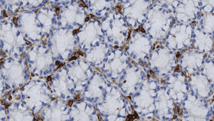
Florence Anquetil-Besnard: For advanced users like me, the support team is also there to bring you even further than what you thought. For example, once I had a big challenge with a complex macrophage staining project involving multiplex techniques. The heterogeneity of the staining was problematic, with intensity varying significantly across samples. In the image analysis community, heterogeneity of fluorescent staining is a nightmare. So, I called the support team, and they guided me through an unconventional use of the software, not something that is taught at first sight. With their help and additional processing steps, we managed to overcome this variability, enabling us to deliver a reliable and robust analysis to our client.
This experience highlights the importance of not only overcoming technical challenges but also of being able to deliver solid, scientific solutions to our clients.
Florence Anquetil-Besnard: In the imaging community, it’s crucial to stay updated and leverage the most advanced tools to remain competitive. Our requirement was for a flexible solution capable of meeting the market’s existing standards. While the Discovery software works for up to 4 markers, the transition to real multiplex analysis requires a more sophisticated approach. The introduction of Phenoplex™ has significantly simplified the analysis of complex multiplex immunostains.
Didier Merciris: Another major advantage that influenced our decision was the exceptional support provided by Visiopharm. As a “newbie” to the software, I really appreciate the constant availability of the support team whenever I need them. They respond quickly and efficiently to any queries or issues, which has been incredibly reassuring.
With Visiopharm, you’re never alone with your challenges; there’s a supportive community, the blog, and a dedicated support team ready to assist.
Visiopharm: What specific features of our software do you find most helpful?
Florence Anquetil-Besnard: With Visiopharm, the process is intuitive: start with an image, add annotations, and if opting for deep learning, proceed to train and validate, among other steps. But what is also really nice is the APP Center – it’s like entering a magical world where you can pick an existing app created by someone else, make minor adjustments to fit your needs, and then all of a sudden you have your own customized app, that you didn’t spend time to create, yet still reliable and efficient.
This flexibility is really helpful to adjust our workflows, offering both time savings and inspiration by exposing us to ideas from other APPs, created by other users in multiple different ways.
Moreover, the software’s versatility extends beyond just brightfield images, but multiplex fluorescent and special stains, enhancing our research scope. We are empowered to combine different histological techniques, like integrating IHC and ISH, allowing simultaneous investigation of proteins and RNA, or merging special stains like H&E or PAS with targeted IHC staining.
We can extract as much information as we want from the minimal amount of tissue, which is very helpful when you don’t have a lot of samples to work with, so we can maximize the analysis with just one image.
Didier Merciris: I used to work with another solution, and to be honest, Visiopharm is faster than the other software. It’s significantly quicker because it can process multiple images concurrently. While the speed can vary based on the user’s IT infrastructure, Visiopharm consistently delivers faster results.
Compared to our former software, we’ve seen a substantial reduction in processing time, enabling us to deliver data to clients much more rapidly. Another advantage is the user experience, the software was built with an accessible interface that is very easy to use after a little training.
Visiopharm: Could you share some insights into how the software has added value to your business and your customers?
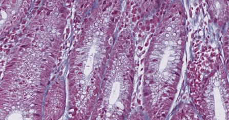
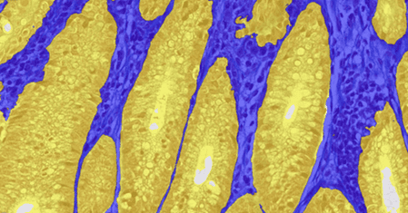
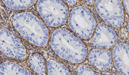
Didier Merciris: As you know, the power of Histology lies in its spatial resolution at the cellular level.
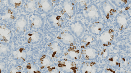
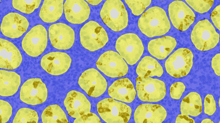
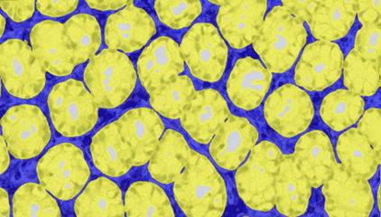
We had a client interested in quantifying mucus production in epithelial cells from a diseased colon, without considering inflammatory cell infiltration.
The AI capabilities of Visiopharm significantly aided us in meeting the client’s needs within an extremely short timeframe.
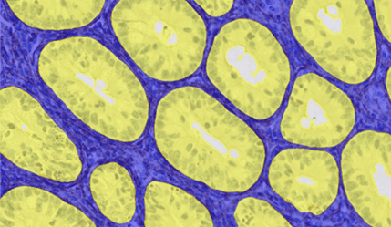
Moreover, thanks to deep learning, we can tailor our algorithm to work with any staining. Now, if there are any color variations on the slide, we can simply add them to the APP.
This allows for much greater consistency in our data, which is a significant advantage for us and our clients.
Florence Anquetil-Besnard: An important aspect is our adaptability as a CRO; it’s not limited to Histology. We can adjust to almost any image. A recent unique project involved working with an image from a camera—not even a microscope, but a camera. We developed a program that automates the counting of tumor nodules in photographs from an ex vivo organ.
Previously, scientists had to manually count all the nodules. We’ve automated this task. By collaborating closely with our colleagues, we’ve enhanced the reproducibility and reliability of organ assessments. This project was internal, but ultimately, our client benefitted the most because we were able to deliver faster results.
Stay Ahead – Realize the potential of AI-driven precision pathology.
In conclusion, NovAliX’s integration of Visiopharm’s image analysis software has significantly transformed their histology and image analysis workflows, offering substantial benefits to their services and clients:
- Enhanced research capabilities: The adoption of deep learning and artificial intelligence within Visiopharm has empowered NovAliX to overcome previous limitations in image analysis, enabling more detailed and accurate data extraction from tissue samples.
- Customization and flexibility: The ability to customize algorithms and adapt to a variety of image types has allowed NovAliX to tailor their services to the specific needs of their clients, enhancing the adaptability and reach of their offerings.
- Overall business value: By leveraging Visiopharm’s technology, NovAliX has not only enhanced their service quality but also solidified their position as a cutting-edge CRO, capable of delivering high-quality, reliable results to their clients in the drug discovery industry.
Curious to hear more about Visiopharm software? Contact us here.
Reach out to NovAliX to hear more about their services here.
Share this article

