Transcript
As I mentioned, we study different types of chronic liver disease, and what we’re doing is looking at the hepatic microenvironment. We have archived liver tissues over thirty years worth, in all different types of chronic liver disease. As I mentioned, steatotic, our fatty liver disease, as well as, different types of emerging infections such as Ebola virus, chronic infections like hepatitis c virus. And so what we’re able to do is go back into this archive liver tissue, and our, r o one funded work is able to go back, look at that tissue microenvironment, and by looking at different signatures at the protein and gene level, trying to see if that liver was going to be an inflammatory liver or a quiet liver.
So no matter what type of injury we have in the liver, we all want to have a very tolerogenic liver. So in other words, if we have some type of insult, whether it’s a medication, a little bit of alcohol, some virus, we do not wanna overreact to that injury. If we do, if we did, we would all have cirrhosis or end stage liver disease. So what we really want is a liver that has proper wound healing.
Just like when we cut ourselves or cut a finger. Right? We want that to heal quickly, not leave a scar. That’s the same thing we want to happen in our liver.
However, well, for example, all all of the antigens from our our gut, go to the liver every day through the portal vein. So if we reacted to every one of those antigens, we would all have cirrhosis. Right? So we need to have the right wound healing response.
And so some patients, for some reason, have more of an inflammatory microenvironment than others. Right? So for example, why do the you could have two patients, both with chronic hepatitis c, same genotype, both females, similar age, same amount of time since they had the virus. One has hardly any scarring in the liver, and one has cirrhosis.
Why is that? And that’s what we’re trying to answer. And we’re not only looking at, you know, we’re looking at all different types of of chronic liver diseases. So today, I’m gonna show you some of, these data.
So here’s the three diseases types or infections I’m gonna focus on. Steatotic liver disease, which now we call this mastoid or mash. It’s a it’s a a long word, but it’s metabolic associated steatotic liver disease. And when you get a lot of inflammation or injury, we call it a metabolic associated steatohepatitis, Ebola virus. We have a a nonhuman primate model that we’ve used here, and receive these tissues again, formal and fixed tissues out of our BSL four laboratory. And I’m also gonna be showing you a little bit how we’ve used Visiopharm in our patients with chronic hepatitis c.
But first, I need to tell you what the normal liver looks like so that when you see injury, it’ll be easy for you to appreciate. So the normal liver, has, here with this marked with this PT, has a portal track area, which has bile ducts. There’s three structures in here, bile ducts, hepatic arteries, and portal veins, and a little bit of collagen around the portal track. In a lot of our diseases, there is a lot of inflammation in this area that spills out into what we call the lobula of the liver, and these are all of our hepatocytes.
And when we have inflammation in these areas, we call that hepatitis.
You can get hepatitis from, like, as I mentioned, steatosis or fatty liver disease, viral infections, for example, immune mediated diseases such as autoimmune hepatitis.
This is the central vein here. Blood flows through the liver towards the central vein, and bile flows out of the liver to the bile duct out into the small intestine. This is a trichrome stain here, which we use to look for the amount of collagen or fibrosis. This is a normal liver here where you see just a little bit of blue in the portal tract area, but you really should not see much blue in the rest of the liver.
So I’m gonna start off with steatosis, our fatty liver disease, but I want you to keep in mind that a lot of our liver injury resides on a lot of these factors. And a lot of them are really, really obvious obvious. Right? So, poor diet, lack of exercise, obesity.
There are some genetic things that can cause steatonic liver disease. Being older over the age of fifty, metabolic syndrome, diabetes, high lipids, and a big one in all liver diseases, not just steatonic or fatty liver disease, it’s the inflammatory microbiome in the in the gut. Right? So if you have a quiet or non inflamed gut, then your liver should be non inflamed. They they are very, very connected.
Also, alcohol overuse, as you know, can cause inflammation in the liver. One of the big ones that can cause inflammation in the liver in our diet is high fructose corn syrup. It’s in, like, everything. Processed food foods can cause inflammatory liver disease, but all of these play a role in developing steatotic liver disease.
So, you know, all of us are you know, you can’t avoid everyone sometimes, but you really wanna try to avoid multiple hits. Right? You don’t wanna have alcohol, poor diet, obesity, diabetes. The more that you have, the more likely you are gonna have a liver that becomes inflamed and pro fibrotic and starts to cause the cascade of causing a scarred liver.
So, I’m giving you example here again in steontotic liver disease because I’m gonna show you those data first. But just keep in mind, this is really our goal. On the left here, I’m showing a happy, nice, healthy liver. This is a liver that kinda turns this yellowy color that has fat in it. Once you some people can have a lot of fat in their liver, and they never progress to getting activation or fibrosis.
So we really wanna keep our livers in this side of this, of this diagram. But But once we start moving into this inflamed liver, these little stellate cells get activated and become myofibroblast, lay down collagen. We get cirrhosis. And once we get cirrhosis, we’re at risk for developing hepatocellular carcinoma or liver cancer.
So these things, again, keep a happy liver, weight loss, even coffee is protective, believe it or not. Exercise, Mediterranean diet is really what we want. Lots lots of veggies, not processed foods. And then the things that are bad for our livers, diabetes, obesity, age, alcohol, and and a lot of lifestyle factors.
Very important to know too that in our patients with steatotic liver disease, a lot of these risk factors also lead for cardiovascular disease.
So how do we do this? So if we look at the liver microenvironment, as I mentioned, we really focus on macrophages. Why are we interested in macrophages? Because Kupffer cells, the main type of macrophage found in the liver, are the the number one non parenchymal cell besides hepatocytes found in our liver.
And it’s their job to kind of sit here on this side. This is the the bloodstream side here inside the endothelial cells of the liver. They sit there kind of like cellular, like, fly fly paper, basically. They kinda sit there and stick there, and they keep with these liver sinusoidal endothelial cells.
They are really the important gatekeepers to keep the liver quiet. Once this these cells are no longer quiet, these endothelial cells allow other cells like circulating macrophages or modify monocytes to enter the liver and start this inflammatory cascade. So once this inflammation starts in the liver, all of these other cells behind the endothelial cells in the space of disc get activated, such as delayed cells, turn into those myofibroblasts, and lay down collagen. And there’s a lot of different markers that we can look at in this and these are the panels that we’ve designed.
I’m gonna show you some of these work today. We’ve looked at pro inflammatory macrophage markers like CD fourteen, markers like CD one sixty three that are important for for m two or fibrosis.
And this MAC two eighty seven marker is really nice nuclear marker to look at these systemic macrophages or infiltrating macrophages. And then we also look a lot of druggable targets, a lot of clinical trials to to help treat fatty liver disease or steatotic liver disease have targeted macrophages.
And so what we’ve done is we really wanna know, does everybody have the same level of these druggable targets in their liver? Are they are we all the same? And we really hypothesized that, in fact, we would be very different. There’d be a lot of heterogeneity.
Here’s just a little bit to show you, some of the trials. These are all of the trials that are in, in, right now being tried to to study and to target macrophages or hepatic’s delayed cells to decrease fibrosis progression in patients with steopathic liver disease. And unfortunately, several of these, these trials ended early because they didn’t meet their endpoints. So what we hypothesized was sorry. What we hypothesized, for example, with the astinocrivarac, which targets CCR two in the liver. You know, these patients all have baseline liver biopsy collected and were given this, Synocrivoroc or CCR two, CCR five antagonist.
After year one, about twenty percent of the patients responded, and everyone was so excited. But then after the end of year two, there was no no really difference in the end of the trial. Well, we hypothesized number one, did all of the patients have a lot of CCR two to be given a CCR two inhibitor? Maybe what happened to the other eighty percent of people that didn’t respond?
Our second question was, at the end of year two, you’ve been inhibiting these cells now for a year. So are they still there? We never we really wanted to get the answer to these questions, but it’s really hard to get your hands on this tissue. So we did these studies on our archive tissue.
In addition, the lectin three, there’s another drug that targets the lectin three. It has a really long name up here. Wanted to to look the level of expression there. And some of the other ones that we are currently optimizing our laboratory are the FXR receptor and the thyroid hormone receptor beta.
Many of you may know Madrigal, this ResMediram, was the first FDA approved drug for treatment of NASH. And so now we’re looking at those targets in the liver. Important difference in this one is this is an agonist. So So when you have liver disease, the expression of this drugable target should go down.
So we’re looking for decreased expression for that.
So just moving on to show you the difference between a couple of patients with spheotonic liver disease. On the left, I’m showing you an example. So I’m a pathologist. These are just two patients that I saw in my microscope.
Just a normal I get these these liver biopsies all the time. We call these patients we call it a activity score, or this is basically the amount of inflammation or injury in the liver. And when we say stage, we’re staging the amount of scarring or the amount of fibrosis in the liver. So when I look in the microscope, patient one over here looks really quiet.
This patient looks like they have a liver that has just a little bit of fat and barely any scarring at all. So this liver patient one in my microscope looked healthier to me.
In contrast, patient two over here had a lot more inflammation, six out of eight inflammatory score, tons of fat. All these holes you’ll notice when I comparing it to that control I showed you, lots of holes in the liver. This is all these are all fat droplets. So tons of inflammation, swelling of the hepatocytes, a very unhappy look at looks very inflamed microenvironment in the microscope and also more fibrosis.
So more blue. Right? More scarring here. So this looked worse in the microscope. And also lots of these macrophages, these hot pink cells here.
These are macrophages, and these are what really prompted me to start my entire research career when I was a a fellow in Pittsburgh about over ten years ago now. I saw these cells all the time, and I just wanted to see them there with a with a, you know, PASD stain or a special stain. I wanted to know their phenotype. Were these eighty one sixty three, were these infiltrating macrophages?
What type of cell were these were these cells? And Visiopharm is what really helped me determine their phenotype. I’m gonna show you that.
So I’m hearing some background noise. I don’t know if I I I’m not hearing anything. Okay. Good. I’m it sounds like it’s coming just to let you know, I just I don’t know if it’s gonna be on the recording or not, but there might be a little background noise.
So here’s the the first paper I’m going to show you. And I put the PMID numbers from all of the papers I’m gonna be showing you, today. This is the first one on steatotic liver disease, and it goes through all of our, methodology, how we did the Visiopharm analysis, what type of, you know, images we use. It has everything step by step in here if you’re interested in in trying to repeat these experiments.
And this is just a, a larger picture of our workflow. So as I mentioned, we have these archived liver biopsies from patients with steatonic liver disease or controls based on these, tissues that we have. We do molecular for half, and we do multiplex staining for the other half. We download the multi component TIFF files, and we analyze them with Visiopharm software using the these files here shown on the right.
So just to show you the difference and and why Visiopharm is so powerful. So these are molecular data. This is the NanoString sprint data. And I just wanna point out a few things. You know, this is homogenizing the entire liver tissue, and and a lot of these different macrofars aren’t gonna be seeing significant differences. But, again, this is completely homogenized liver, and you can see that we didn’t see significant differences when we looked here. However, when you go down to these druggable targets, CCR two, Galectin three, and these other ligands for these as well as CD163.
We started to see really, really significant differences in controls.
Patients like patient one with mild disease and then patients that have, severe liver disease. And the really interesting thing that we saw was within cirrhosis was we had marked heterogeneity, meaning there’s a lot of differences. Each one of these dots here is an individual patient for CCR two and the lectin three. Why would we wanna give these patients down here?
You can see why would we wanna give these people a CCR two inhibitor when they had their expression was no different than a control? So in other words, maybe these people didn’t respond to your drug because they don’t have a lot of CCR two. However, the patients at the high expression at the top highly significantly different than controls. So these are the patients we might want to do more precision medicine and give the group that a higher expression of the drug and not all the patients.
I think we would have seen more efficacy or more a higher percent of patients responding. Had we done a precision medicine approach with a lot of these drugs in clinical trials?
And now how do we go and look at these patients on an individual level? And that’s where Visiopharm comes in. So now I’m showing you the hepatic microenvironment. I’m showing you, just representative images from a normal liver, a patient with mild steatohepatitis, a patient with, moderately active and then severely active steatohepatitis.
And Visiopharm allows us to segment the tissue into the portal track areas. You can see here separating the portal track from the lobule and again here a portal track from lobule and then we can go and analyze these different cell types in these different regions. We’re looking at CD one sixty three target, CCR two, as I mentioned, the lectin three, and then the the systemic macrophages marked by MAC three eighty seven.
And we’re able to generate these data by looking at the different expression of these markers in the microenvironment using Visiopharm software. So quite interestingly, what we we did not expect, we we thought we would see heterogeneity. That that we expected because as we know, humans are very, very different. But interestingly, patient one that had that really quiet liver on in my microscope actually had a lot higher expression of CCR two than patient two that looked like it had a really inflamed.
So, again, patient one was probably going the wrong direction with a high CCR two expression. We probably didn’t wanna give patient two a a CCR two inhibitor. Maybe something else would have been better for this patient. And again, the lectin three was much higher in that patient that appeared to have a quiet liver than inpatient two.
So again, you know, precision medicine is really going to be where we need to go to have the most success treating these different types of liver disease.
Again, that I showed you steatotic liver disease, but we also found when we looked at this is these are all Visiopharm data. So if you look, this is our multispectral imaging. Our phenotype markers here for macrophages on the left, and then tip our tissue segmentation using Visiopharm separating it into portal tracks and mobiles. So the way that we design this, it’s really your application for separating originally. I think now there’s been a lot of software updates, but originally this was for basically separating tumor and non tumor, and we were able to use this algorithm to separate portal tracks from from lobules. So that’s how we separated the different microenvironment.
And then using the different phenotype mapping, this is with Visiopharm. So now we’re taking our multiplex, and making it where we can see every phenotype and where it is in the hepatic microenvironment. So just remember, every single dot in these images is a different phenotype. The gray ones are null, meaning they didn’t up any of our antibodies, but you can see controls have a very, very a lot of this yellow phenotype, very, very quiet on our tSNE plot, meaning not a lot of diversity in cell types cells that are very, very tightly clustered. But as you introduce diseases, here, hep c, here, fatty liver disease, here, autoimmune hepatitis, you increase the number of of different phenotypes and the different complexity of cells.
HCV here, a steatonic liver disease here, and we were really, really surprised to see all of the phenotypes and autoimmune hepatitis. And so now we’re surprised to see all of the phenotypes and autoimmune hepatitis. And so now we’re studying this disease as well. And all of these Disney plots were generated with Visiopharm.
The cool thing that we’ve been able to do now over the last couple of years is now we know when we look at the colors of phenotypes in the microenvironment and the tisnies, we can use our our phenotype profile map to look at the different patients and the different expression of these different phenotypes. So as I told you, this patient here had a lot of this kinda lime green phenotype. I can tell you that’s a lot of CD fourteen. I can look the the controls have a lot of CD fourteen.
In contrast, these hot pink cells here that we see a lot of these in autoimmune hepatitis, I can come and tell you all the phenotypes lobules and autoimmune. So it’s really, really powerful. I can literally go and say, oh, wow. There’s a lot of these hot pink ones on the portal tracks in autoimmune.
There’s a lot of these yellow ones in the lobules and controls, and this is what Visiopharm is allowed us to do.
So the next disease type, I’m gonna tell you, some more did you have any questions first? Leticia, I don’t know if you wanted to ask any questions about the daniptotic liver disease so far.
I think it’s it was a really clear presentation.
You can go on. If you need to drink so hard, by the way. I’m actually good. I I only have a few more slides. So we’ll do Ebola virus, which is only a few slides, and then we’ll go on and show you the hep c work.
Cool. Okay. So next, I’m gonna talk about, you know, we were working with my MD PhD student, Timothy Waniger, and this is a recent paper that we had published in Frontiers in Immunology. It just came out the last couple of weeks, actually, last month.
So we’re really excited about this work. So as I mentioned to you, we started to see okay. We have these macrophages and all the different, you know, human liver diseases, and we’ve seen a lot of similarities. So, basically, injury in the liver causes inflammation and and a lot of diseases that causes infiltration of these systemic or CCR two expression expression, expressing macrophages.
Well, could we see those cells in a totally different disease? Right? There all the work out there right now, we see it in steatotic or fatty liver disease, but what about Ebola virus infection?
I mean, totally different. Right? Can be fatal, more of an acute inflammatory disease. But you know what?
Macrophages have to be important. I told you there’s a lot of them in the liver. We know there’s injury in the liver. We get a lot of macrophages.
There’s already been other work that that macrophages expand during disease. So can we use FFPE or formalin fixed tissues? This time, given from other investigators, we’re already doing this work. They already have the tissue blocks.
So we had we we got the control, Ebola tissue blocks from our primary research center here in Texas. And then we were able to get, from doctor Jason Comer, we were able to get, some liver tissue and lung tissue from nonhuman primates for this work.
So what did Visiopharm allow us to do here? So we wanted to determine in these infected NHPs the differences in different macrophage phenotypes in the hepatic microenvironment. So we use Visiopharm to quantify the percent population between our uninfected nonhuman primates and our infected, primates with Ebola. I mean, this is really powerful. Right? Not not a lot of people have these tissues, and we were able to be able to actually quantify the different numbers of phenotypes using Visio.
Next, we also looked at, as I mentioned to you, there’s been so much work on CCR two and steatotic liver disease. But what about Ebola? Totally different. Right?
An emerging infection, really, really more of an acute liver disease can be fatal in animals wanting to look at expression. And very interestingly, there are drugs to target. I just showed you. There are drugs to target CCR two, and we found by using Visiopharm, and we actually also used an antigen from Ebola VP thirty five.
So we made a multiplex panel with, dapyard nuclear stain, macrophage, for Cooper cells, the infiltrating macrophages, mac three eighty seven and CCR two, and the Ebola and, antigen. And we were able to use Visiopharm to calculate the different, percent expression here. So CCR two, we didn’t see any significant difference. However, in this Mac three eighty seven population, we saw a lot of these infiltrating in the liver, and of course, a lot more Ebola virus antigen in the infected animals than uninfected.
So, again, you know, very, very powerful to look at the different levels of expression, in these animals.
Here, moving on to showing you the difference in, hepatitis c virus. I’m just gonna show you some examples here in chronic infection. So I just showed you how we did that in acute infection, and now we’re gonna show you some work that we’ve done in chronic infection to viral hepatitis c. So for this study, it’s actually, from my post doc, doctor Daniel Milligan, who is a pathology resident.
He just successfully, defended his PhD, work back in September. So this is all of his work. What we did was we went back to our archive tissues, and we took, liver biopsies from patients before they got treated with hepatitis c, and then we collected liver biopsies after they were treated with hepatitis c. So what we’re doing is we’re looking at the liver microenvironment and control patients without disease, patients before when they still had active HCV infections, and then after their liver microenvironment was cleared to the virus to to see the differences in the immune response before and after treatment, with these new direct acting antivirals for hepatitis c.
And these these work are just a beautiful, images that we have from Visiopharm. So here on the left, I’m just showing you the normal h and e’s. So the control liver is very similar to that one that I showed you earlier. We or the liver’s quiet without any inflammation and then showing you before treatment.
And some of these patients had persistent inflammation after treatment, and then some patients had no inflammation after treatment. And, again, showing you the different phenotypes in the microenvironment, you can see very, very quiet livers and controls versus active viral infections, people with persistent inflammation in their livers after treatment, and then more of a quiet liver after treatment. And we made all these tizzies again using Visiopharm. You can see a lot of the gray are quiet or just normal cells and controls, and the people that had quiet livers after treatment, they really look like controls.
But the peep people with active infections and the people with persistent inflammation after sheema look very, very similar, but with some different phenotypes. And the cool thing is is, again, like I mentioned, you can actually go hone in on these different cell types and figure out with our, phenotype profile maps of what type of phenotypes these are. So this is really, really, interesting. These these are actually using our t cell panel.
So we looked at c d three, c d four, c d eight, memory t cells, and regulatory t cells with FOX b three. And so it’s really, really cool to see that we had a lot of memory cells in the patients after treatment and a lot of these different, really in that patient’s post treatment, the inflammation you’re seeing is a memory response. So it’s really cool. We were able to determine that, and a lot of it is actually in the portal track as you can see, not in the labial.
So a lot of different things we were able to tell using Visiopharm and chronic hepatitis c.
Here’s just an example. Looking at the expression of these different phenotypes using the phenotype profile maps, and we’re showing you here these different, again, memory really high expression of memory phenotypes in these, livers and patients after treatment.
So in summary, this just summarize all of the different things. You know, today, I talked a little bit about these different disease types. First, I showed you steatotic or or fatty liver disease. I showed you a little bit of our work in, an emerging infection, Ebola virus disease, as well as looking in a chronic viral infection like hep c. But in summary, what we’re really hoping to start doing, you know, we get liver biopsies here. I get liver biopsies almost every day as a liver pathologist.
Right now, pathologists just give us a diagnosis and a fibrosis stage, but we still have precious liver tissue on that tissue block. If I was a patient getting a liver biopsy, I wanna know as much as I possibly can from that precious liver tissue that I gave you. So in other words, why can’t I give you more information? That’s the way we need to move.
I can tell you just like a colonoscopy. Right? You go you go, you get a biopsy. The doctor says, oh, your your colonoscopy was normal.
See you in ten years. Oh, no. We found something. We found a polyp. We found a precancerous lesion.
We need to come back more frequently so we prevent disease. Why can’t we do that with your liver? There’s no reason we can’t. Right?
We do it already with cancer. So when a clinician orders a liver biopsy, the liver biopsy is sent to pathology, I’ll still give you the diagnosis. I’ll still give you the amount of inflammation in my microscope. But I will also take that precious liver tissue, and I will look at protein expression and gene expression to give you the more more information about your liver.
So this is what we all want. We all want a happy liver. Your liver looks like this. You know what?
Miss Johnson, your liver’s quiet. I think you’re doing good. You’re just keep doing what you’re doing. I’d like to see you back with your annual physical.
We’ll check your liver enzymes. You’re good to go. Oh, no, mister mister Smith. Your liver is quite inflamed.
It has a lot of pro inflammatory signatures. It has a lot of fibrosis. Even though it looks okay in the microscope now, I’m not happy with how your liver looks. You need to watch your alcohol, watch your diet, lose some weight.
Maybe we can add some of these these new GLP one agonists like Ozepic or Zapatide. Right? We can talk about some of these emerging, drugs for macrophages. Your liver is not happy.
I need to see you more frequently. You need more precise follow-up. So this is really where we’re going, precision medicine and personalized care. And honestly, you know, Visiopharm is the type of software that’s gonna get us there.
And so I really hope today that I’ve shown you, a lot of the work that we’re doing and convinced you how cool and, all of these emerging things that we’re gonna be seeing coming out in the next few few years. And with that, this is my my research team. I have to thank my group because everyone in this picture has done a lot of work. These are just some of the, some of the people that have done the work.
This is Tim Weniger. He’s my MD PhD student. He’s applying for internal medicine residency now, so he’s back in clinic. This is doctor Daniel Milligan.
He is a pathology resident who just finished his PhD. He’s from Puerto Rico. This is doctor Omar Salarayaga.
He is my main collaborator and and also helps run my lab. He’s done so much of this work. I’ve worked with him for over seven years. This is doctor Esteban Oroyabe.
He’s my research scientist. He’s also from Columbia. He’s done a lot of the immune mediated and autoimmune work. This is Joe Gosnell.
He is a pathology resident doing GI liveries, getting ready to go to Mayo for his fellowship. And, of course, I I had to show my boys here, Jude and Ryder, as well. And I think that’s it for, for now for the presentation.
The liver’s microenvironment holds the key to understanding and addressing complex liver diseases, from fibrosis to emerging infections. In this lecture, Prof. Stevenson-Lerner shares her experience leveraging Visiopharm software to explore the liver microenvironment, offering unique insights into macrophage-mediated immune responses and the use of advanced AI tools for spatial biology analysis. She also highlights how Visiopharm empowers her team to study liver diseases with unprecedented precision, ensuring reproducible and impactful research outcomes.
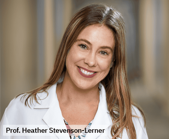
Prof. Heather Stevenson-Lerner
Heather Stevenson-Lerner, MD, PhD, is a Professor in the Department of Pathology, Division of Surgical Pathology at the University of Texas Medical Branch. Her clinical focus includes liver, transplantation, and gastrointestinal pathology.