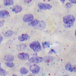
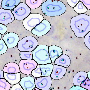
Nuclei surrounded by intermediate and strong NT positive staining.
#10120
Hepatocellular carcinoma (HCC) is a primary cancer of the liver and a leading cause of cancer-related death worldwide, see [1]. Important risk factors include chronic liver disease and cirrhosis, and the incidence of HCC is expected to continue to escalate. HCC is an aggressive cancer and early diagnosis is critical for the survival of patients. In situ hybridization (ISH) is used for the detection of markers of HCC. Presented in 2012, RNAscope is one of the more recent RNA in situ hybridization techniques that allows visualization of multiple target genes. RNAscope uses a probe design strategy that amplifies the signal and suppresses the background, which results in increased sensitivity and specificity, see [2].
The APP works on dual probe RNAscope images. Cells and probe signals are automatically detected and cells are classified according to the number of probes associated with the cell. Both probe signals and cells are quantified.
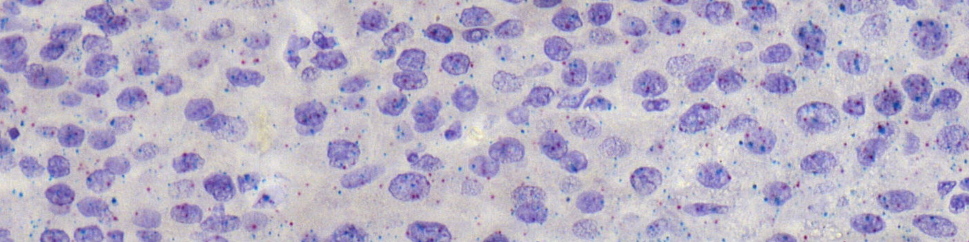
Nuclei surrounded by intermediate and strong NT positive staining.
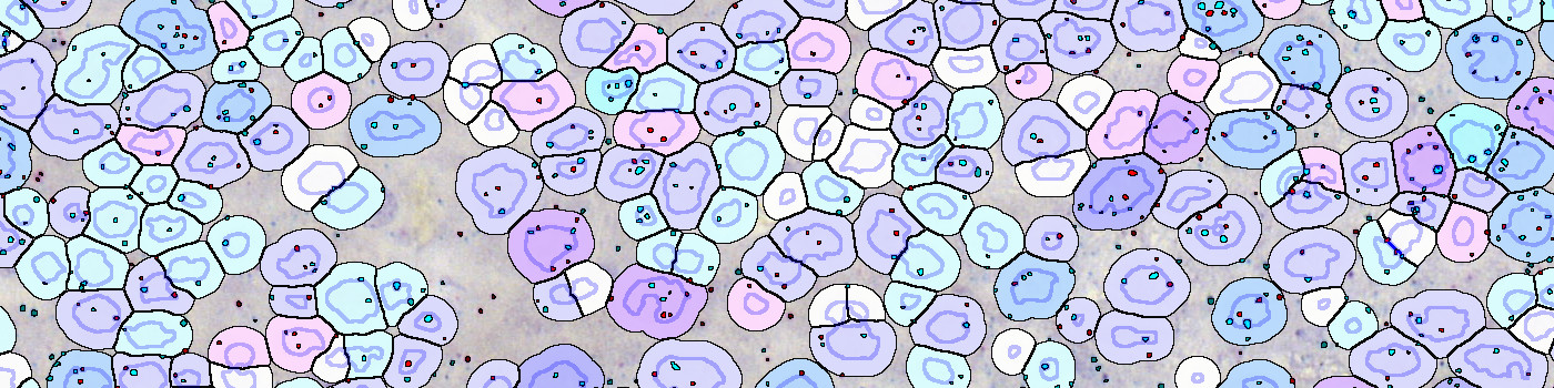
Analyzed image showing identified nuclei (blue) and simulated cytoplasm (yellow).
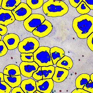
Analyzed image showing cells classified based on contained probes.
Quantitative Output variables
The output variables obtained from this protocol are:
Workflow
Step 1: Manually outline one or multiple ROIs in an image
Step 2: Load and run the APP “10120 – RNAscope, Hepatocellular Carcinoma” for the quantification of cells and probe signals
Methods
Initially, nuclei and signals from two different probes (red and teal) are detected inside a manually outlined region of interest (ROI). Both nucleus and probe signal detection use a polynomial blob filter to enhance their features. The nucleus detection is based on a combination of the red, green, and hematoxylin color-deconvolution bands, whereas the detection of red and teal probe signals are based on the eosin and chromaticity red color-deconvolution bands. Large areas of probe signal are separated into smaller sized probe signals. Cytoplasm is simulated by dilating nucleus areas until another cell or white space is reached, or until a maximum cell diameter of 4 µm is reached (see FIGURE 2). Probe signals are further classified as coming from inside a nucleus, inside cytoplasm, or outside cells. Probe signal count and area are quantified for each class. Cells (nuclei + cytoplasm) are classified based on the number of red and teal probe signals coming from each cell (see FIGURE 3). All cell classes are quantified.
Staining Protocol
RNAscope® 2.5 LS Duplex Assay (Advanced Cell Diagnostics, Inc.)
Keywords
RNAscope, dual probe, liver, cancer, digital pathology, image analysis, in situ hybridization, ISH, hepatocellular carcinoma, HCC
References