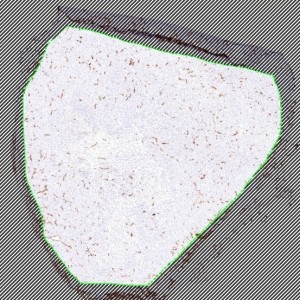
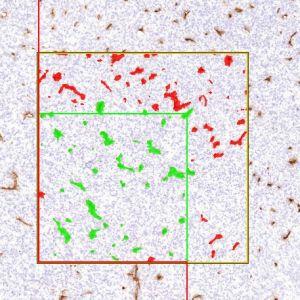
Outlining the ROI.
#10022
Angiogenesis, or neovascularization, is the formation of new blood vessels originating from the endothelium of existing vasculature. Angiogenesis is known to play an important role in the neoplastic progression leading to metastasis. CD31 immunohistochemistry (IHC), is widely used in experimental studies quantifying tumor neovascularization in immunocompromised animal models implanted with transformed human cell lines.
By using this APP, neovascularization can be quantified from full virtual slides.
Outlining the ROI.
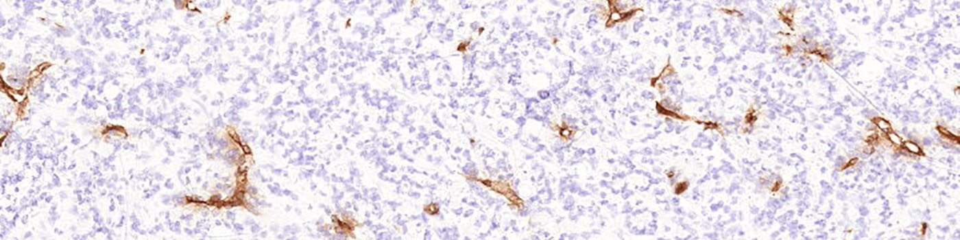
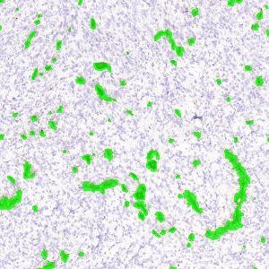
One field of view of the original image at 2X (scaled down to fit this space).
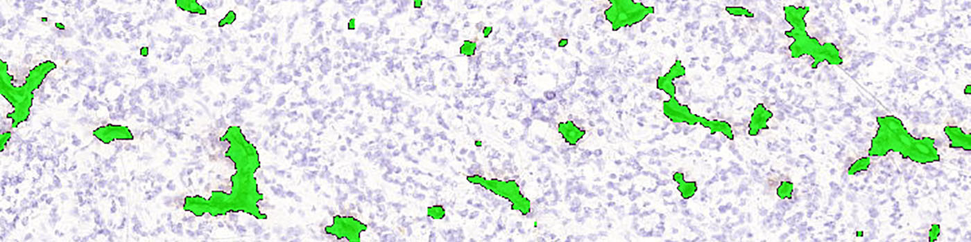
Classification of the vessels. Note that the lumen of the vessels has been filled in order to get the cross-sectional area measurements for the vessel profiles.
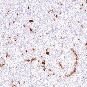
An un-biased counting frame is included to ensure that each vessel is counted only once.
Quantitative Output variables
Angiogenesis is often quantified as the cross-sectional vessel area fraction and the microvessel density within the region of interest.
The outputs from this protocol are therefore:
Methods
All vessel profiles are identified within a region of interest (ROI), which can either be outlined manually (see FIGURE 1) or by an automated method based on image analysis.
NOTE ON AREA MEASUREMENTS
Only the vessel walls are stained, but the analysis protocol is automatically filling the lumen, in order to get the cross-sectional area measurements for the vessel profiles (see FIGURE 3).
NOTE ON COUNTING
Analysis of full virtual slides takes place in a tile-by-tile fashion. If not handled appropriately, vessel profiles that are intersecting with neighboring tile boundaries would be counted twice (or more). Using an unbiased counting frame, see [1], this can be avoided (see FIGURE 4). This principle is implemented in the present APP. Depending on vessel size and density, the application of this principle could make an important difference.
Staining Protocol
There is no staining protocol available.
Keywords
Angiogenesis, neovascularization, CD31, microvessel density, cross-sectional area fraction, quantitative, digital pathology, image analysis.
References