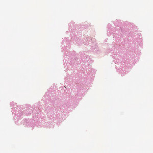
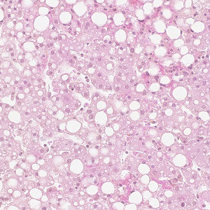
H&E-stained liver tissue.
#10119
Liver steatosis or Fatty liver disease (FLD) is a disease that is normally associated with alcoholism or obesity in humans. This reversible condition is an abnormal retention of fat within the hepatocytes and results in an accumulation of triglyceride within the hepatocytes. Macrovesicular steatosis is the most common form of steatosis, where large droplets containing triglycerides displace the nuclei to the periphery. Often macrovesicular steatosis can be present with both large droplets (ld-MaS) and small droplets (sd-MaS), that grow together into a mass.
This protocol can be used to quantify fat within hepatocytes in H&E stained liver sections. The APP detects the total area of liver tissue and quantifies the fat inside the tissue. The identified fat droplets are separated into small-droplets (sd-MaS) and large-droplets (ld-MaS). In the end, the total fat percentage and the fat percentage of each of the two droplet types are given.
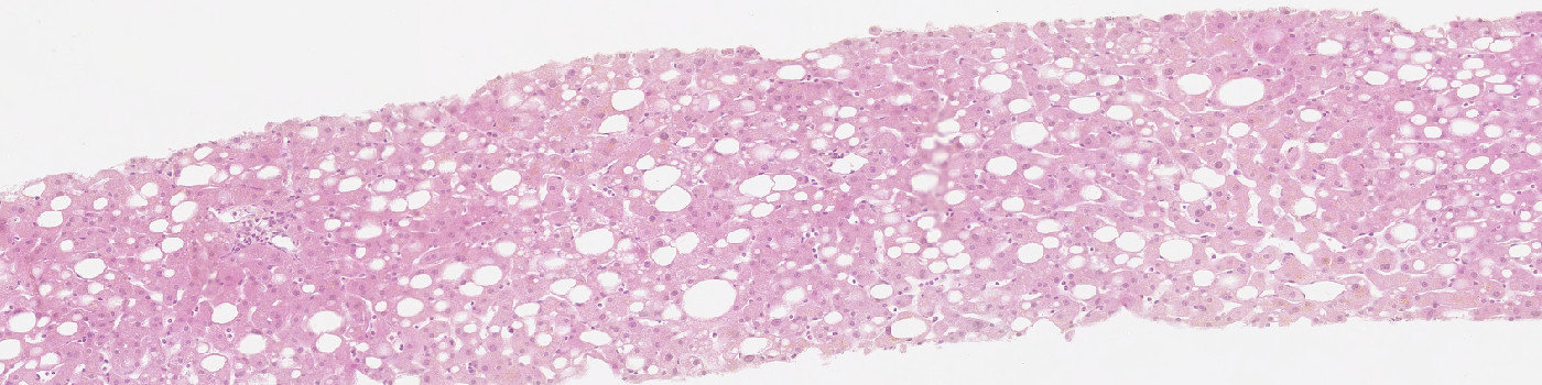
H&E-stained liver tissue.
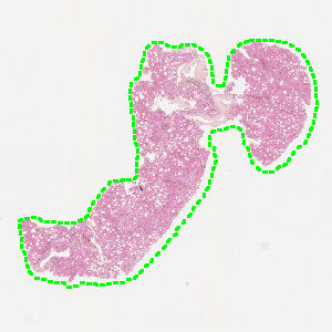
Result of analysis with APP: “01 ROI Detect”. The liver tissue is automatically outlined.
Close up of fatty liver tissue.
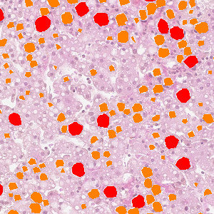
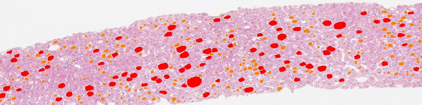
The accumulation of fat inside the liver tissue is identified. The red labels represent large-droplet macrovesicular steatosis (ld-MaS) and the orange labels represent small-droplet macrovesicular steatosis (sd-MaS).
Auxiliary APPs
APP: “01 ROI Detect”
This APP can be used for automatic and precise identification of tissue present on image.
Quantitative Output variables
The output variables obtained from this protocol include:
Workflow
Step 1: Load the APP for tissue detection “01 ROI Detect” which outlines the liver tissue in the image.
Step 2: Load the quantification protocol “02 Analyze” which identifies the fat within the hepatocytes and calculates the fat percentages.
Methods
The first image processing step involves a segmentation of the liver tissue (see FIGURE 2). Afterwards the droplets inside the liver tissue are identified. Based on the shape and size of the droplets they are identified as either small-droplets (sd-MaS) or large-droplets (ld-MaS). By looking at the form factor structures that are too elongated are discarded as being droplets (see FIGURE 4).
Staining Protocol
There is no staining protocol available.
Keywords
Liver steatosis, fatty liver disease, small-droplet, large-droplet, HE, H&E, quantitative, digital pathology, image analysis.
References
LITERATURE
There are currently no references.