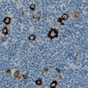
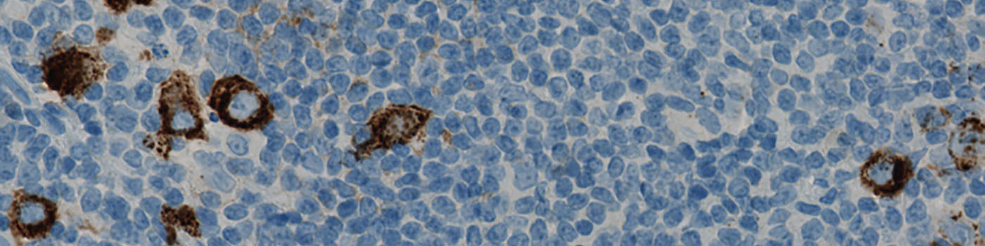
Original image.
#10052
Hodgkin’s disease, is a type of lymphoma, and is also referred to as Hodgkin’s lymphoma. The disease will spread from one lymph node group to another in an organized manner. During the advanced disease stages, systemic symptoms will usually develop. A common histopathologic finding is multinucleated Reed–Sternberg cells, which are the neoplastic cells of Hodgkin’s lymphoma.
CD15 is useful for the differential diagnosis of Hodgkin disease vs. non-Hodgkin lymphomas and for classification of leukemia. The ratio of CD15 positive cells is an indicator for the progression and aggression of the disease. In some neoplastic diseases like Hodgkin disease, myeloid leukemia and gliomas, CD15 expression is inversely correlated to dedifferentiation and progression.
In CD15 positive cells the antigen expression may be membranous, diffuse cytoplasmic or related to the Golgi zone and is expressed on most terminally differentiated myeloid cells (granulocytes, monocytes/histiocytes and Langerhans cells).
Here, CD15 is heavily expressed in the cytoplasm of the neutrophil granulocytes, making it difficult to see the nucleus of the cells when the chromogen is filling the cytoplasm. CD15 is more moderately expressed in the neoplastic Hodgkin and Reed-Sternberg cells. The cell and nucleus diameter of these cells are also often far larger than the diameter of the neutrophil granulocytes. Therefore, the Hodgkin and Reed-Sternberg cells will appear as cells with a (negatively stained) large nucleus or multi nuclei surrounded by brown cytoplasm, where the staining is less dense than for the neutrophil granulocytes. The cell area of a neoplastic cell will usually be at least 400 µm.
Smaller, single nucleus cells, with intense staining (or where the nucleus is not visible due to the density of the chromogen) will most likely be neutrophil granulocytes.
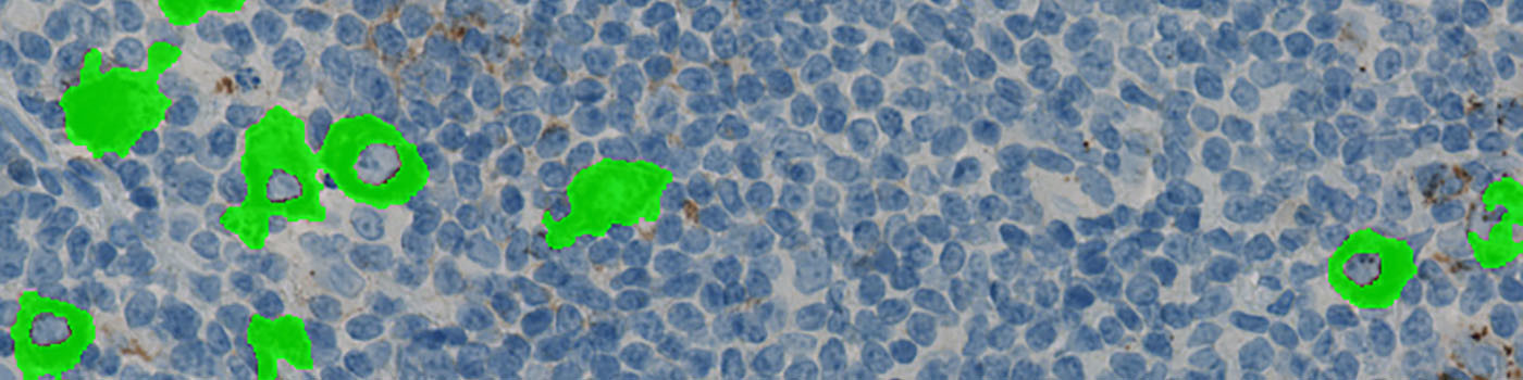
The purpose of the APP is to locate and segment the number and area of the neoplastic cells, as well as determine the positive ratio compared to the total analyzed area.
Original image.
Classified image.
Negative nuclei are removed if they are too far away from the Hodgkin’s Lymphoma cells.
The negative nuclei are used to verify that the Hodgkin’s Lymphoma detections are not false positives and afterwards removed.
Quantitative Output variables
Methods
METHODS
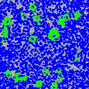
The first processing of the APP is the nuclei detection, which involves a segmentation of all negative (blue) nuclei and positive cells (brown) within the ROI (which is defined by the user) (see FIGURE 2). Nuclei are identified by assigning a label probability to all pixels in the image, resulting in a label probability image. The label probability image is found by an intensity dictionary – a dictionary with small image patches. The intensity dictionary can be coupled to a label dictionary from which the label probability image is obtained. Based on this image, segmentation of cell/nuclei can be done by choosing the most probable label in each pixel, see [1]. A method for nuclei separation which is based on shape, size and nuclei probability is used, employing a fully automated watershed-based nuclei segmentation technique. The method is an extension of the method proposed by Jung and Kim, see [2], where an h-minima transform is used before applying the watershed.
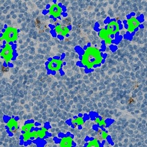
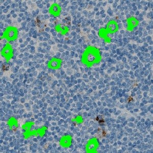
Thereafter, all negative nuclei that are not near a Hodgkin’s Lymphoma are removed, as well as large clusters of nuclei (see FIGURE 3). The remaining nuclei are used to verify that the green labels cells are actually Hodgkin’s Lymphoma cells, which yields the final result (see FIGURE 4).
The APP requires a manually drawn ROI. The APP further requires that the ROI does not include areas with moderately large amount of neutrophilic granulocytes since they resemble the Hodgkin Lymphoma in such a degree that they will disturb the algorithm.
Staining Protocol
There is no staining protocol available.
Additional information
This APP was developed in cooperation with Professor Mogens Vyberg from NordiQC and Aalborg University Hospital.
Keywords
Hodgkin’s lymphoma, CD15, expression, intensity, tumor, positive area ratio, area, number
References
USERS
This APP was developed in cooperation with Professor Mogens Vyberg from NordiQC and Aalborg University Hospital.
LITERATURE
1. Dahl, A. L. et. al., Learning Dictionaries of Discriminative Image Patches, British Machine Vision Conference 2011, DOI
2. Jung, C. et. al., Segmenting Clustered Nuclei Using H-minima Transform-Based Marker Extraction and Contour Parameterization, IEEE Transactions on Biomedical Engineering 2010, 57 (10), 2600-2604, DOI
3. Arber, D.A. et. al. CD15: A Review, Applied Immunohistochemistry 1993, 1 (1), 17-30, DOI