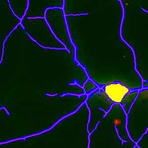
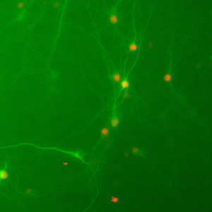
20X view of the cell culture (scaled down to fit this space)
#10050
The effects of progressive neural tissue degeneration can be imitated by administering compounds based on neuro-toxic viral proteins to neuron cultures. The toxins cause oxidative stress damage and free radical production, and will trigger apoptotic cell death in the neurons.
The capability and the power of a given therapeutic agent in reducing the effects of degeneration and death of neuronal tissue can be quantified by screening the health status of the neuron cultures, previously treated with toxins, after the administration of such therapeutic agent. Further, it can be evaluated whether the therapeutic agent has axo-dendritic protective effect, meaning the capability to minimize pruning of axons and dendrites for every neuron.
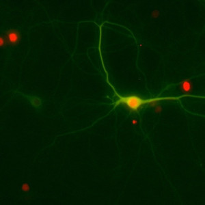
The images here were acquired with fluorescence microscopy, and the cell cultures are from rat mixed hippocampal cultures which were transfected with a neuron-specific promoter containing the red fluorescent protein tdTomato, with Tau fusion protein linked to the tdTomato. These have been generated with double transfection-yellow making the neurites appear in green, while the cell bodies appear red. Toxins, along with a therapeutic agent are added to the culture to study the effect on the cell body number and process length.
20X view of the cell culture (scaled down to fit this space)
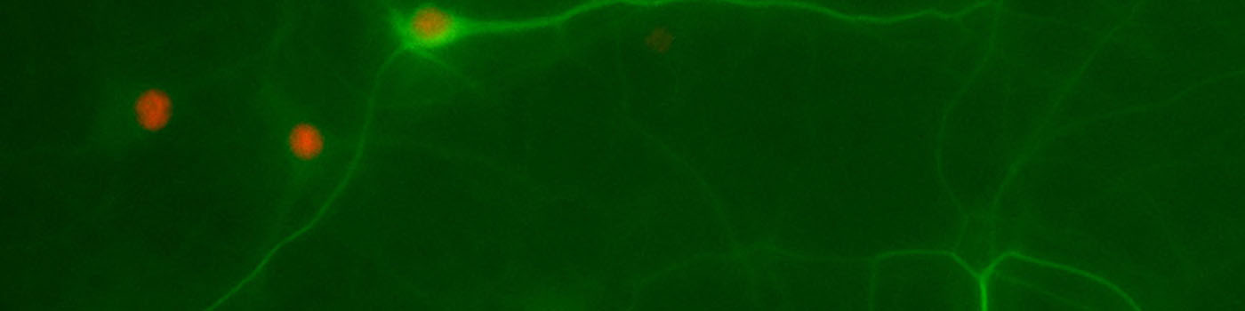
20X view of the cell culture (scaled down to fit this space). Notice the difference in background illumination compared to FIGURE 1.
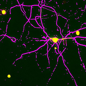
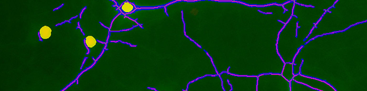
Initial classification of the same area as shown in FIGURE 1. The sensitivity is set so that as many neurites as possible are identified. This in turns detects a relatively high degree of background noise.
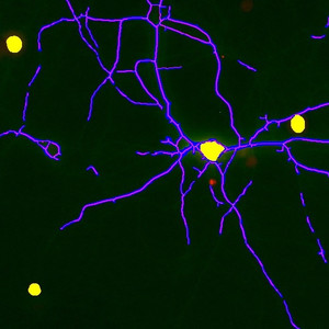
During post processing falsely detected cell bodies and neurites are removed, and the neurite area is skeletonized to facilitate the calculation of the process lengths.
Quantitative Output variables
The output variables obtained from this protocol include:
Methods
There is a relatively large variation of the background in the images , which is corrected for during pre-processing. Further, a polynomial line filter is applied to enhance neurites. With the correct pre-processing steps, a robust detection of both neurites and cell bodies is obtained with a simple threshold classifier. The settings are adjusted to a high sensitivity to pick up neurites with low signal intensity, for the initial classification.
During post processing background noise resembling neurite segments (small, scattered objects) are removed along with falsely detected cell bodies. Post processing also includes a step to “skeletonize’ the identified neurite area to facilitate the calculation of the process length. The resulting segmentation after post processing is shown under Examples.
Keywords
Cell culture, neural tissue degeneration, axo-dendritic protective effect, neuro-toxicity, fluorescence microscopy, quantitative, digital pathology, image analysis.