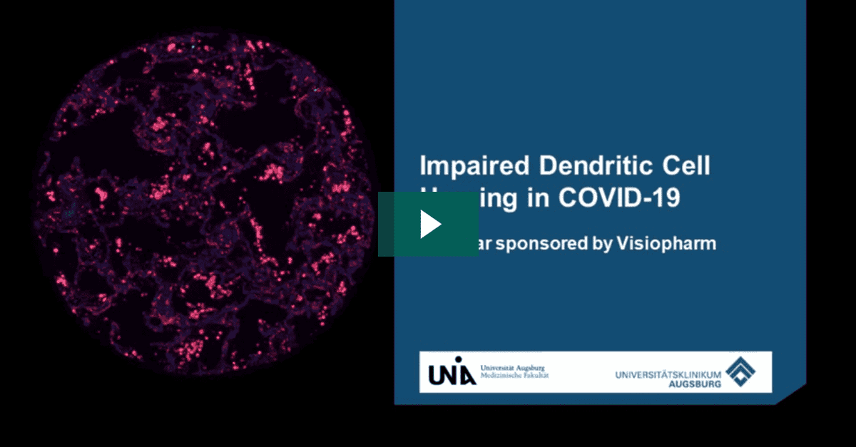
The high mortality rate of COVID-19 is largely due to acute respiratory distress syndrome (ARDS), which is characterized by diffuse alveolar damage (DAD). Additionally, severe cases of COVID-19 often result in a cytokine storm and a disrupted adaptive immune response. Most previous studies on this issue have focused on the peripheral cell count and the functionality of immune cells.
The team at UK Augsburg in Germany used multiplexed immunofluorescence to study the impact of SARS-CoV-2 on antigen-presenting cells. Like MERS-CoV and SARS-CoV, SARS-CoV-2 appears to impair the maturation of dendritic cells (DCs), which is characterized by a switch in surface antigen expression. This switch enables the cells to travel to lymph nodes and activate T-cells.
To shed light on the local inflammatory infiltrate, the team compared the cell populations of professional antigen-presenting cells (APCs) in the lungs of COVID-19 autopsy cases in various stages of DAD. They found an increased number of myeloid dendritic cells (mDCs) in later stages, but with no significant upregulation of maturation markers in DAD specimens with high viral load. This accumulation of immature mDCs, which are unable to reach lymph nodes, results in an inadequate T-cell response.
Overall, this study highlights the need for further research to understand the impact of SARS-CoV-2 on the immune system and to develop more effective treatments for COVID-19Discover how the team employed digital analysis of multiplex IF images to gain insights into the unique immune profile in fatal COVID-19 cases. Investigate the potential impacts of SARS-CoV-2 and related coronaviridae on antigen-presenting cells. Learn about the role played by the UKA in COVID-19 autopsies.
-
- Experience how the team used digital analysis of multiplex IF images to learn more about the specific immune landscape in fatal COVID cases
-
- Explore the possible effects of SARS-CoV-2 and related coronaviridae on antigen presenting cells
-
- Understand the role of the UKA in COVID-19 autopsies
Prof Ralf Huss, University Hospital Augsburg
Ralf Huss, MD, PhD, is a Professor of Pathology and the Managing Deputy Director of Pathology and Molecular Diagnostics at the University Hospital in Augsburg, Germany. He is also the head of the Center for Digital Medicine.
Huss holds board certifications in anatomical, experimental, and molecular pathology and boasts over 30 years of expertise in histopathology, immunology, cancer research, and oncology.

Lukas Borcherding, Medical Student, Technical University of Munich
Lukas Borcherding is a trained nurse who has been pursuing a degree in medicine at the Technical University of Munich, Germany since 2015. In 2020-2021, he took a hiatus from his studies to concentrate on the COVID-19 pandemic and examine the unique immune profile in fatal cases. Following the completion of his project, Lukas has resumed his studies and is presently finishing the practical component of his education. Upon receiving his license, he intends to work in internal medicine.
Join Fluidigm and Visiopharm at the Imaging Mass Cytometry Summit for a demonstrtaion of the uitility of Visiopharm’s Phenotyping and Deep Learning toolbox for advanced multiplex phenotype analysis of Hyperion Imaging System multiplexed data sets.
We will provide an overview of Visiopharm’s flexible and intuitive Deep Learning and Advanced Phenotyping solutions to provide a comprehensive analysis of highly multiplexed IMC datasets. Specifically, we will demonstrate Visiopharm’s approach to and scope of cell segmentation and phenotyping algortithm development using real world IMC examples.
-
- Tissue mining to enhance anlysis
-
- Cell segmentation by deep learning
-
- Robust multiplex phenotyping
-
- Integration supported phenotyping
Brit Boehmer, Account Executive, Sales US, Visiopharm
Brit Boehmer is an Account Executive for the US West based in Denver, CO. He has a master’s and Ph.D. in physiology from Oklahoma State University and completed postdoctoral research in reproduction, nutrition and fetal growth. Brit joined Visiopharm in 2020 and supports clients with pre-sale APP development and advice on image analysis and histopathology workflows.
-
- Discover how the Visiopharm platform can be used for multiplex analysis
-
- Review an example analysis of an mIF 8plex image
-
- Learn how the new Visiopharm Data Insights tool can be used for exploring image objects
-
- See an example analysis of a Fluidigm Hyperion CyTOF image covering tissue and cell segmentation using AI deep learning, phenotyping and an example tumor microenvironment region analysis

Dr Fabian Schneider, PhD, Service Project Lead, Visiopharm
Dr Fabian Schneider is part of Visiopharm’s R&D and Product Management team, responsible for phenotyping products as well as service projects for custom APP development. Fabian has over 10 years of international experience in cancer biology and immuno-oncology, working in academic research labs, clinical research teams and computational pathology groups in both academia and biopharma. Fabian received his Dr phil. nat. in Cell Biology in 2011 from the Johan Wolfgang Goethe University Frankfurt, Germany.
Quantitative histological analyses are used to infer pathological diagnoses and identify biological drug targets in human and laboratory animal samples. Each histological sample often include millions of cells imposing a need for automated unbiased quantitative analysis to replace manual assessment.
We have trained a convolutional neural network that can identify individual nuclei inferred from DAPI fluorescence, allowing automated quantitative analysis of various fluorescent marker levels in nuclei. We used this convolutional network to assess proliferation from nuclear Ki67 fluorescence and apoptotic DNA fragmentation from terminal deoxynucleotidyl transferase dUTP nick end labelling (TUNEL) at a single cell level in human kidney biopsies. Upon retraining, the convolutional neural network was adaptable to identify nuclei from pancreas emphasizing the versatility of the network. Taken together, these results demonstrate an AI-based automated method for quantifying fluorescence levels of various markers in diverse tissues such as kidney and pancreas.
Tobias Højgaard Dovmark, Research Scientist, Novo Nordisk
As a research scientist at Novo Nordisk, Tobias Højgaard work with image analysis of histological samples. He has previously worked for Visiopharm as an image analysis specialist. He has a PhD in Physiology, Anatomy and Genetics from University of Oxford and an M.Sc. in Molecular Biomedicine from the University of Copenhagen.
Dr Fabian Schneider, PhD, Service Project Lead, Visiopharm
Dr Fabian Schneider is part of Visiopharm’s R&D and Product Management team, responsible for phenotyping products as well as service projects for custom APP development. Fabian has over 10 years of international experience in cancer biology and immuno-oncology, working in academic research labs, clinical research teams and computational pathology groups in both academia and biopharma. Fabian received his Dr phil. nat. in Cell Biology in 2011 from the Johan Wolfgang Goethe University Frankfurt, Germany.
Immunotherapy has transformed the treatment of metastatic and recurrent solid tumors but is challenging in that only a minority of patients respond. Therapies that rely on immune activation, such as checkpoint inhibitors, have been shown to be especially difficult due to the complex and heterogeneous immune escape mechanisms which can develop in each patient. Therefore, development of robust biomarkers coupled with spatial analysis of tissue is key for enabling rational patient selection and the design of precise combination therapies.
The use of multiplex immunohistochemistry/immunofluorescence (mIHC/IF) provides much needed insight into cellular composition, cellular functions, and cell-cell interactions. Importantly, recent studies have used mIHC/IF to explore specific immune cells as part of the tumor immune microenvironment (TME) and found that it is helpful for clinical prognosis and efficacy prediction in patients with cancer.
In this presentation we will show a streamlined unique workflow supporting whole slide imaging of an 8-plex mIF and H&E fusion on a single tissue slide for a comprehensive tissue immunophenotyping analysis.
-
- The utility of a high throughput, high-plex (Immuno-8) staining and mIF assay development for scientists and clinicians
-
- Demonstrate how advanced AI-driven image analysis can be applied to discover cell types, populations and morphological context
-
- Discuss how whole slide image analysis of the tumor microenvironment can provide insight into specific cancer types
Fabian Schneider, PhD, Service Project Lead, Visiopharm
Dr. Fabian Schneider is part of Visiopharm’s R&D and Product Management team, responsible for phenotyping products as well as service projects for custom APP development. Fabian has over 10 years of international experience in cancer biology and immuno-oncology, working in academic research labs, clinical research teams and computational pathology groups in both academia and biopharma. Fabian received his Dr phil. nat. in Cell Biology in 2011 from the Johan Wolfgang Goethe University Frankfurt, Germany.

Angela Vasaturo, PhD
Associate Director, Biomarker Strategy and Applications