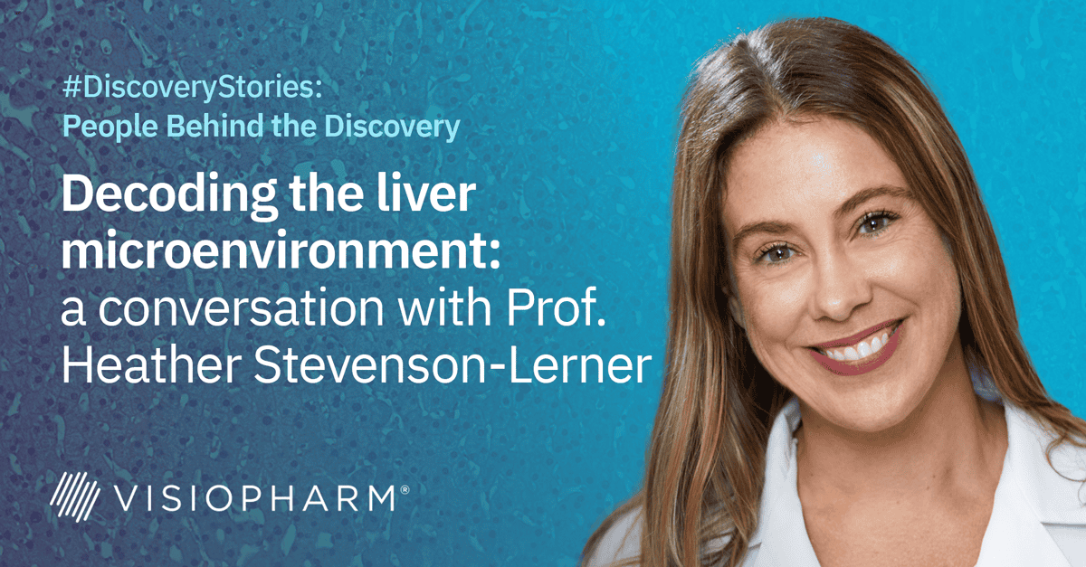
The liver’s microenvironment holds the key to understanding and addressing complex liver diseases, from fibrosis to emerging infections. In this lecture, Prof. Stevenson-Lerner shares her experience leveraging Visiopharm software to explore the liver microenvironment, offering unique insights into macrophage-mediated immune responses and the use of advanced AI tools for spatial biology analysis. She also highlights how Visiopharm empowers her team to study liver diseases with unprecedented precision, ensuring reproducible and impactful research outcomes.

Prof. Heather Stevenson-Lerner
Heather Stevenson-Lerner, MD, PhD, is a Professor in the Department of Pathology, Division of Surgical Pathology at the University of Texas Medical Branch. Her clinical focus includes liver, transplantation, and gastrointestinal pathology.
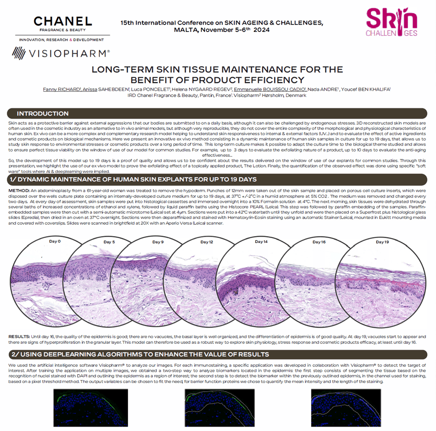
Skin acts as a protective barrier against external aggressions that our bodies are submitted to on a daily basis, although it can also be challenged by endogenous stresses. 3D reconstructed skin models are often used in the cosmetic industry as an alternative to in vivo animal models, but although very reproducible, they do not cover the entire complexity of the morphological and physiological characteristics of human skin. Ex vivo can be a more complex and complementary research model helping to understand skin responsiveness to internal & external factors (UV .. .) and to evaluate the effect of active ingredients and cosmetic products on biological mechanisms. Here we present an innovative ex vivo method consisting in a dynamic maintenance of human skin samples in culture for up to 19 days, that allows us to study skin response to environmental stresses or cosmetic products over a long period of time. This long-term culture makes it possible to adapt the culture time to the biological theme studied and allows to ensure perfect tissue viability on the window of use of our model for common studies. For example, up to 3 days to evaluate the exfoliating nature of a product, up to 10 days to evaluate the anti-aging effectiveness …
So, the development of this model up to 19 days is a proof of quality and allows us to be confident about the results delivered on the window of use of our explants for common studies. Through this presentation, we highlight the use of our ex vivo model to prove the exfoliating effect of a topically applied product, The Lotion. Finally, the quantification of the observed effect was done using specific “soft ware” tools where Al & deeplearning were implied.
Fanny Richard1, Anissa Sahebdeen1, Luca Poncelet1, Helena Nygaard Regev2, Emmanuelle Bouissou Cadio1, Nada Andre1, Youcef Ben Khalifa1
- IRD Chanel Fragrance & Beauty, Pantin, France
- Visiopharm Hørsholm, Denmark
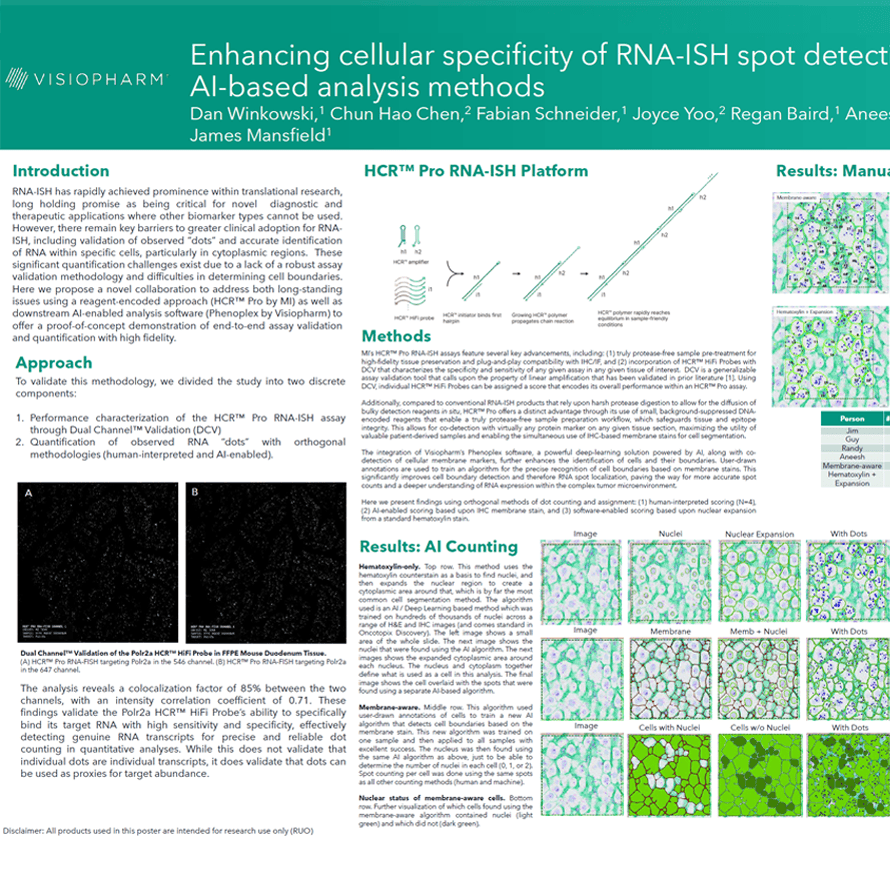
RNA-ISH has rapidly achieved prominence within translational research, long holding promise as being critical for novel diagnostic and therapeutic applications where other biomarker types cannot be used. However, there remain key barriers to greater clinical adoption for RNAISH, including validation of observed ”dots” and accurate identification of RNA within specific cells, particularly in cytoplasmic regions. These significant quantification challenges exist due to a lack of a robust assay validation methodology and difficulties in determining cell boundaries. Here we propose a novel collaboration to address both long-standing issues using a reagent-encoded approach (HCR™ Pro by MI) as well as downstream AI-enabled analysis software (Phenoplex by Visiopharm) to offer a proof-of-concept demonstration of end-to-end assay validation and quantification with high fidelity.
Dan Winkowski1, Chun Hao Chen2, Fabian Schneider1, Joyce Yoo2, Regan Baird1, James Mansfield1
- Visiopharm A/S, Horsholm, Denmark
- Molecular Instruments, Los Angeles, CA, USA
 Bettina Winkler
Categories:
25665
Innovative Imaging Analysis Approaches: The PCKD mouse model as a key source for human ADPKD understanding
Bettina Winkler
Categories:
25665
Innovative Imaging Analysis Approaches: The PCKD mouse model as a key source for human ADPKD understanding
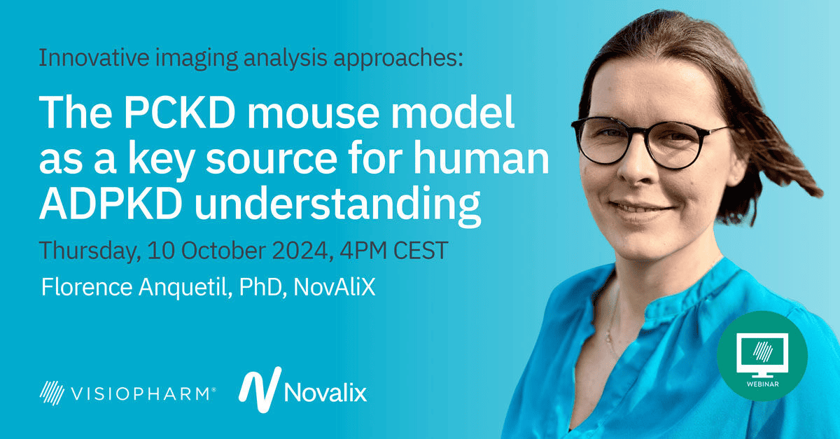
Autosomal dominant polycystic kidney disease (ADPKD) is an inherited systemic disorder mainly associated with mutations in the PKD1 gene and characterized by the development of multiple cysts in the kidneys and other organs. There is still a high unmet need for treatment options for patients with ADPKD because Tolvaptan, the only approved treatment, has limited efficacy and non-negligible side effects.
To mimic naturally occurring human PKD1 mutations, a Pkd1 inducible knockout mice strain was established at Novalix. Embark on an exciting journey as we explore the histological characterization of the PDK1flox/flox model using cutting-edge Visiopharm technology. We will unveil captivating insights into the disease’s origins and evolution, revealing the intricate structural changes that define this strain. Join us as we uncover the hidden stories within the tissues, paving the way for potential breakthroughs in understanding and treatment!

Florence Anquetil-Besnard. PhD
Florence Anquetil-Besnard is a Histology Project Manager at NovAliX. After a PharmD and a PhD in Immunology, she began her career in the autoimmune field (rheumatoid arthritis, diabetes) and specialized in histopathology and quantitative image analysis. She is a passionate advocate for digital pathology and loves to solve challenging projects using artificial intelligence (AI) tools. Florence joined the Galapagos team in 2020 to implement the newly formed kidney disease histology area. Her current work within NovAliX also includes oncology projects in diverse organs (brain, pancreas, liver, xenograft tumor…).
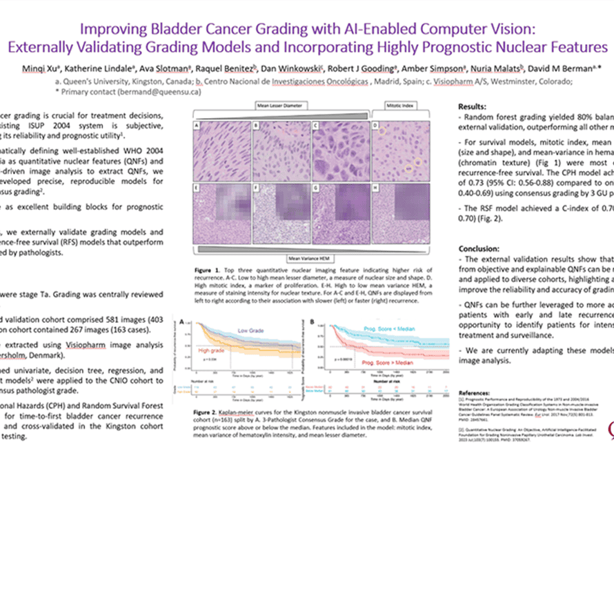
- Bladder cancer grading is crucial for treatment decisions, but the existing ISUP 2004 system is subjective, compromising its reliability and prognostic utility.
- By mathematically defining well-established WHO 2004 grading criteria as quantitative nuclear features (QNFs) and employing AI-driven image analysis to extract QNFs, we previously developed precise, reproducible models for expert consensus grading.
- QNFs serve as excellent building blocks for prognostic classifiers.
- Using QNFs, we externally validate grading models and create recurrence-free survival (RFS) models that outperform grades assigned by pathologists.
Minqi Xua1, Katherine Lindalea1, Ava Slotmana1, Raquel Benitezb2, Dan Winkowskic3, Robert J Goodinga1, Amber Simpsona1, Nuria Malatsb2, David M Bermana1.
- Queen’s University, Kingston, Canada
- Centro Nacional de Investigaciones Oncológicas , Madrid, Spain
- Visiopharm A/S, Westminster, Colorado;
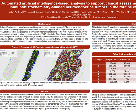
Cell proliferation is commonly assessed to grade neuroendocrine tumors (NETs) and aid in the evaluation of patient prognosis and treatment selection. To grade NETs, a proliferation index (PI) is evaluated based on the presence of immunohistochemical staining of the Ki-67 nuclear antigen. In the gastrointestinal tract, grading is performed using WHO criteria for PI as Grade 1-3 (less than 3%, 3%-20%, and greater than 20% respectively). It is recommended to count at least 500 cells followed by calculating the percentage of Ki-67 positive nuclei for cases with the Ki-67 index close to grade cut-offs on eyeballing. Ki 67 proliferation index near cut-offs is a real challenge for practicing pathologists.
Vidya Arole MD1*, Jesse Kulewsky1, Jenifer Caldara2, Rachel Scarl DO1, Swati Satturwar MD1, Zaibo Li MD1, Wei Chen MD1, Anil Parwani MD1, Giovanni Lujan MD1
- The Ohio State University Wexner Medical Center
- Visiopharm Corporation