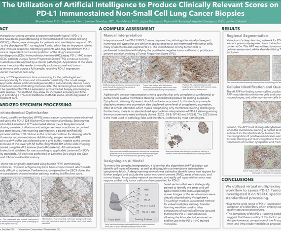
Immunotherapies targeting complex programmed death-ligand 1 (PD-L1) interactions have been groundbreaking in the treatment of non-small cell lung cancer (NSCLC), offering new strategies for patients who are likely to respond. PDL1 binds to the checkpoint PD-1 to regulate T cells, which has an important role in boosting the immune response. Identifying patients who may benefit from PD-L1 focused care is dependent on the interpretation of the drug’s associated companion diagnostic (CDx) immunohistochemistry (IHC) assay. PD-L1 IHC assays stratify NSCLC patients using a Tumor Proportion Score (TPS), a manual scoring paradigm which must be applied by a clinical pathologist. Application of the score is complex as it requires the reader to visually exclude stromal and tumorinfiltrating immune cells across a full sample, retaining PD-L1 expression information for true tumor cells only.
The intricacy of TPS application is time consuming for the pathologist and introduces opportunity for inter- and intra-reader variability. Our novel image analysis approach utilizes artificial intelligence (AI) to automatically denote tumor nests and exclude tumor-infiltrating immune cells. The remaining true tumor cells may then be quantified for PD-L1 expression across the full biopsy, producing a TPS for each sample. This method may allow for increased accuracy and timeefficiency in providing a TPS over traditional methods alone, which may result in improved patient care.
Bhavika Patel, PhD1, Stephanie Allen1, Sameer Talwalkar, MD1, Navi Mehra, PhD1, Jeppe Thagaard2, Thomas W. Ramsing2, Agnete Overgaard, PhD2, Jenifer Caldara2
-
- Lanterne Dx, Boulder CO.
- Visiopharm Corporation
Dive into the world of HER2 expression with Visiopharm’s Chief Diagnostics Officer, Dirk Vossen and Technical Sales Specialist, Jeni Caldara. They’ll be showcasing our innovative two-step solution for interpretation. Confirm your HER2 staining consistency for accurate assessment, and use the vendor agnostic HER2Connect™ app for scoring interpretation. HER2Connect™ has a proven track-record in identifying HER2-low status specimens, a traditionally challenging group to identify through manual interpretation. Don’t miss this opportunity to improve your HER2 interpretation process.
-
- Investigate staining proficiency and discover the only commercial AI technology available to monitor HER2 staining consistency continuously
-
- Learn about our IVDR-certified HER2 Breast Cancer APP and see real-world performance data produced at a leading US hospital during a research study
-
- Discover how AI-powered decision support can aid in identifying HER2-low specimens

Dirk Vossen, Chief Diagnostics Officer, Visiopharm
Dirk Vossen, leads a team to develop diagnostic and clinical applications of digital pathology. He has a strong track record of creating value through innovation in digital and computational pathology, covering the full range of development from ideation to validation and certification of medical devices and commercialization strategies. With over a decade of experience as a global leader in building digital pathology solutions, Dirk has expertise in developing whole slide image scanners, image management systems, computational pathology applications, and executing clinical programs. He holds a Ph.D. from the AMOLF Institute and Utrecht University.
Jeni Caldara, Technical Sales Specialist, Visiopharm
Jeni Caldara is an innovative digital pathology professional with 6 years of experience in AI-based image analysis and workflow implementation. With a background in cell biology, she brings creativity to the generation of AI solutions that highlight tissue-specific processes. As a Technical Sales Specialist at Visiopharm, Jeni is based in the Colorado office, where she focuses on translating product offerings and integrating them into existing systems at customer sites.
Join Brit and Brenna from Visiopharm as they delve into our comprehensive AI toolbox for histopathology image assessment. They will begin by discussing the implementation of deep learning algorithms for maximizing data from multiplex phenotypic analysis. Discover how Visiopharm’s ease of use and robustness make it the ideal solution for even the toughest challenges.
-
- Simplifying deep learning algorithms for complex feature analysis
-
- Iterative approach to enhance the success of deep learning applications
-
- Maximizing workflow efficiency and accuracy through APP sequences and integrations
-
- Optimizing applications to tackle analytical challenges
-
- Providing platform-agnostic solutions for multiplex phenotyping data sets
-
- Practical benefits of using digital histopathology workflows
Brit Boehmer, Account Executive, Sales US, Visiopharm
Brit Boehmer is an Account Executive for the US West based in Denver, CO. He has a master’s and Ph.D. in physiology from Oklahoma State University and completed postdoctoral research in reproduction, nutrition and fetal growth. Brit joined Visiopharm in 2020 and supports clients with pre-sale APP development and advice on image analysis and histopathology workflows.
Brenna O’Neill, Technical Sales Specialist, Sales US, Visiopharm
Brenna O’Neill is the Technical Sales Specialist for the US West, based in Visiopharm’s Westminster, CO office. She has a master’s in ecology and over 10 years of imaging experience across multiple platforms, including 3 years in image analysis. As a Technical Sales Specialist, Brenna supports clients with software demonstrations and pre-sale APP development to address their image analysis needs.
Easy-to-use AI-based deep learning is now integrated as the standard analysis methodology in our best-in-class analysis software. Designed for anyone, it allows tissue-based researchers to tackle both simple and complex datasets across a wide variety of applications. In the past, only image analysis experts could analyse complex tissues.
Our new analysis packages include powerful pre-trained nuclei segmentation algorithms suitable for brightfield and fluorescence applications which can be further tuned for even more specificity, and deep-learning-based tissue segmentation to find tissues of interest, exclude artifacts, and enable scoring/counting within specific tissue compartments. Now, anyone with the understanding of tissue morphology can train an AI-based deep learning network to get accurate and reproducible data, making it easy to generate reliable quantitative results needed for breakthrough discoveries and publications.
-
- How the use of deep learning improves the analysis of digital pathology images
-
- The use of deep learning to segment tissues, find artifacts, and localize scoring/counting to specific regions of tissue
-
- Using pre-trained deep learning networks to segment nuclei across a range of tissue and staining types

David Mason, Senior Technical Specialist, Visiopharm
Dave Mason is a senior technical specialist in image analysis, supporting Visiopharm’s UK and European sales team. He has a background in cell biology and microbiology and has spent over a decade in academia, specializing in light microscopy and digital image analysis.

Dr Fabian Schneider, PhD, Product Manager Research, Visiopharm
Dr Fabian Schneider is part of Visiopharm’s R&D and Product Management team, responsible for phenotyping products as well as service projects for custom APP development. Fabian has over 10 years of international experience in cancer biology and immuno-oncology, working in academic research labs, clinical research teams and computational pathology groups in both academia and biopharma. Fabian received his Dr phil. nat. in Cell Biology in 2011 from the Johan Wolfgang Goethe University Frankfurt, Germany.
Artificial Intelligence (AI) continues to serve as a valuable tool for innovative pathologists and scientists looking to extract quantitative tissue data. AI APPs empower readers with another data source by highlighting features of interest, including tissue compartments, tumor volume, biomarker positivity, hot spots of cell populations, and more. During this workshop, Dr Giovanni Lujan, the Associate Director of Digital and computational pathology at the OSU Wexner Medical Center and OSU James Comprehensive Cancer Center, will describe The Ohio State University’s adaptation of a fully digital workflow, highlighting their work with Visiopharm’s AI-based software and APPs.
To provide additional technical application details, Jeni Caldara, Visiopharm’s Translational Technical Sales Specialist, will describe the algorithms and workflows underway at The Ohio State University. Jeni will dive into endpoint specifications, time studies, and share a pathologist’s use case. This workshop will conclude with a brief Q&A session.
-
- Hear from an expert pathologist on their experience with fully digital workflow and how they are teaming up with AI
-
- Learn more about several of Visiopharm’s Translational APPs, including technical, performance, and analytical validation details
-
- Experience a real-world use case for implementing AI-based pathologist support
Jeni Caldara, Technical Sales Specialist, Translational
Jeni Caldara is an innovative digital pathology professional that has spent the past 6 years specializing in AI-based image analysis and workflow implementation. With an education in cell biology, she enjoys generating creative AI solutions which highlight tissue-specific processes. Currently, Jeni is a Technical Sales Specialist at Visiopharm and is based out of the Colorado office, where she focuses on translational product offerings and incorporation into existing systems at user sites.

Dr Giovanni Lujan, Associate Director, Computational Pathology
Dr Lujan is an associate professor of pathology at the Ohio State University, College of Medicine, a clinical gastrointestinal pathologist and the associate director of Digital and computational pathology at the OSU Wexner Medical Center and OSU James Comprehensive Cancer Center. He is board certified in anatomic and clinical pathology with expertise in gastrointestinal pathology and over 15 years of experience in that field.
He completed a surgical pathology fellowship at The Johns Hopkins Medical Institution after graduating from the pathology residency program at The University of Texas Southwestern Medical Center. Dr Lujan has held previous academic appointments at The Johns Hopkins University School of Medicine and The University of Texas Southwestern Medical School.
 Sheila Hansen
Categories:
19314
From high background to complex staining patterns: Using AI to extract what the eyes can see but the software typically cannot tell
Sheila Hansen
Categories:
19314
From high background to complex staining patterns: Using AI to extract what the eyes can see but the software typically cannot tell

Joseph Allison, MSci, Research Technician | UK Dementia Research Institute, King’s College London
Joseph Allison MSci, is a Research Technician at the UK Dementia Research Institute, King’s College London. He studied and received his MSci degree from King’s College London with two extended research projects involving behavioural assays in Drosophila to elucidate the development of ‘negative’ associative learning as well as AAV stereotaxic injections in mice to determine the neural connectivity underpinning sunlight-mediated mood and cognition. In 2019, he joined the Christopher Shaw Lab with the team’s research focus centred around gene therapy and pathomechanisms in amyotrophic lateral sclerosis and frontotemporal dementia.
Over the last two years he has worked in the histology team processing animal tissue for immunohistochemical and fluorescent staining with subsequent imaging on a slide scanner or confocal, multiphoton, and calcium imaging microscopes. He is now in charge of quantifying the images obtained using software such as Visiopharm to develop apps informing business decisions through analysis of efficacy and toxicity.