Q&A: HistologiX discusses their journey with Visiopharm’s image analysis solution.
Today, we delve into a conversation with HistologiX, a prominent UK-based CRO that has adopted Visiopharm software to supercharge its image analysis capabilities and add value to its customers. HistologiX specializes in histology and immunohistochemistry services for pharma and biotech. Recently, their digital pathology team has been on a quest for a software solution that enhances their offering and optimizes their capabilities. Let’s learn more about their journey to find the ideal image analysis solution, the hurdles they once faced, and the enhanced offerings they can now present to their customers
James Clay is the Digital Pathology Manager at HistologiX where he developed their digital pathology capabilities, integrating whole slide scanning with quantitative image analysis. He began his scientific career in diagnostic histopathology, specializing in immunohistochemistry and routine histopathology. After a move to contract research, he took lead roles in the design, conduct and reporting of regulatory (GLP) Tissue cross-reactivity studies with novel therapeutic antibodies.

Connor McCracken is a Research Scientist at HistologiX within the Digital Pathology team. Connor’s primary role is in Image Analysis, using AI-driven software to deliver quantitative endpoints for a range of projects, from tissue morphometry to multiplex immunofluorescence. Connor has a 1st Class Bachelors in Biology with a focus on bioinformatics, and is currently undertaking a part-time Masters in Computer Science.
Visiopharm: Welcome, James and Connor. Can you tell us about HistologiX, your services, and how you differentiate yourself from other CROs?
James Clay: We are a UK-based CRO, heavily concentrating on preclinical and developmental studies. From routine toxicology to complex assays like chromogenic and multiplex fluorescence. We do quite a lot of tissue cross-reactivity studies of therapeutic monoclonal and novel antibody constructs like nanobodies for regulatory submissions, and FDA drug submissions. In the last couple of years, we’ve been focusing much more heavily on immunofluorescence and multiplexing. We have a lot of fourplex assays up and running as well as Ultivue’s Immuno8 panel and have recently been doing studies where we are multiplexing RNAScope with IHC immunofluorescence.
Connor McCracken: There’s been a big demand from clients for us to use image analysis on their studies to gain as much information as possible. We are offering it as a service for most studies, especially the ones where the images are quite complex and there’s lots of data to generate.
James Clay: Our distinction lies in our bespoke client-focused approach. Answering the specific questions and challenges the clients have, rather than offering a big bucket list of our services. We get heavily involved with the study design, concepts, data outputs, and questions that they have. It’s always a two-way approach to working with the clients.
Visiopharm: What problems were you trying to solve and what was the limitation you experienced?
Connor McCracken: The main thing we were also missing was flexibility with the platform. With the solutions we had before, the modules are built in a way that “it’s for this or it’s for that”, whereas we were getting questions from clients that required a very specific approach that was not possible with what we had. We couldn’t answer some of the questions we were getting asked from clients unless we had the full open tool set available to us. For instance, in one study, we collected muscle biopsies and had to segment these muscles into individual fibers and then detect RNAscope signals within them, but also a membrane-based protein marker. This tailored analysis was previously impossible because we couldn’t connect these different analyses.
James Clay: We already had analysis capabilities with existing platforms, but one of the major things we lacked was AI/deep learning capabilities, which feed heavily into ROI (region of interest) segmentation, mining the images as much as possible, increasing the reliability of nuclear detections, and things like that. That was one of the main areas we were lacking in our current toolsets. We were always heavily dependent on excessive amounts of manual annotation, which is very time-consuming.
“After trying multiple providers, Visiopharm stood out with its intuitive design and efficient workflows.”
– James Clay, HistologiX
Visiopharm: How did Visiopharm’s software fulfill your needs? Which features were most relevant for you?
Connor McCracken: Having flexibility now with Visiopharm in the way that you can build your own apps, either using one of the prebuilt ones or building it from scratch and then chaining them together in a very nice and logical way – that’s the kind of thing we were missing before. Being able to go under the hood of algorithms is what we wanted and what we’ve got with Visiopharm’s Oncotopix Discovery. We can now just tell our clients “give us your question and we will figure something out, that meets it specifically.” Now we are able to offer something bespoke, that fits whatever questions our clients have.
James Clay: Primarily, just that ability to go beyond the pre-canned algorithms and develop things that were much more biologically relevant to the questions that our customers were asking. Offering individual solutions, which aligns very well with the way we like to work with the clients.
Connor McCracken: Also, the ease of training and applying deep learning has been really great. It’s super easy to use because there’s the pre-trained nuclear segmentation, which works nicely, but it’s easy to go and add extra training regions. Moreover, if we’re building the deep learning from scratch, it’s a similar workflow and we’ve been really impressed with how quick and easy it has been to work with it. We were concerned that it would be hard to work with it and would take lots of training, but it’s been very easy.
James Clay: After trying multiple providers, Visiopharm stood out with its intuitive design and efficient workflows.
Connor McCracken: Additionally, it kind of extends beyond just the software itself, but the support is really good. The training we’ve had from the academy team has been really good and really in-depth. And now, we just go to them with questions, and they just work through it with us and that’s been really helpful. A first-rate service really.
“With Visiopharm’s AI, we can now offer more in-depth and histologically relevant data, more structures, and functional regions of interest.”
– Connor McCracken, HistologiX
Visiopharm: How does Visiopharm’s software impact the way you now work with clients?
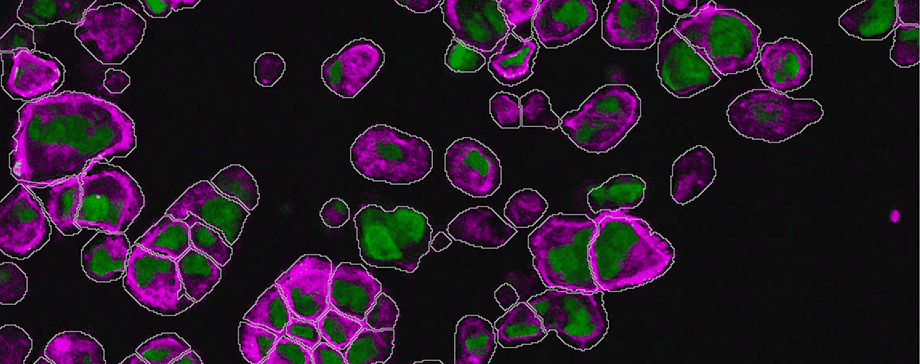
Connor McCracken: There are studies where everything, from tissue segmentation to nuclear detection benefits from having AI. Highly accurate nuclear segmentation can really increase the integrity of the data, especially for complex fluorescent samples. With Visiopharm’s AI, we can now offer more in-depth and histologically relevant data, more structures, and functional regions of interest. Moreover, our client’s needs are evolving, they want more complex questions to be answered through images rather than just a very general quantification. Now, we can offer that.
Another study we had was a multiplex IF with a variety of immune and macrophage markers in a xenograft model. We had planned to use Pan-Cytokeratin as a tumor marker, but realized after staining, that 50-75% of the tumor had differentiated to a state, that it was no longer expressing PanCK.Before Visiopharm, this would have meant weeks of manual annotations.
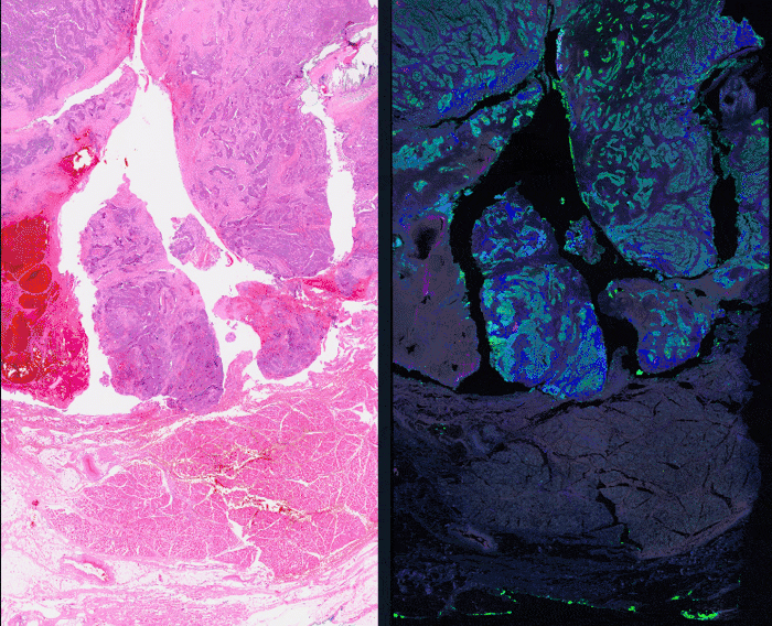
But now, we just used the Tissuealign feature to align the IF and the H&E image. Using deep learning to find the tumor allowed us to exclude other tissue regions like muscle, so we ended up with better information than we would have had just using PanCK. After detection, the regions transferred nicely onto the fluorescent slide, so that we were then able to quantify and phenotype the cells in the tumor/stroma compartments more accurately.
“Before we were always having to fit the study design to the analysis capabilities, whereas now we can fit the analysis approach to the requirements of the study.”
– James Clay, HistologiX
James Clay: This refined methodology has wide applications. We’ve begun exploring its use in multiplex fluorescence. By integrating registered H&E in the same section or adding other fluorophores, we can provide more cellular histological context. This level of detail, especially when combined with deep learning, enables more refined regions-of-interest analysis and a higher data granularity. We are doing many more studies now in the spatial biology sphere, like immune-oncology with lots of multiplex markers. The ability to build the spatial relationships how we want, not how it is pre-canned in an algorithm already, is really helpful. Visiopharm has transformed our approach. Not only does it enable deeper morphometric analysis, but it also allows for more precise quantification of protein expression within detected objects. One of the driving factors for our switch was the software’s ability to offer in-depth customized filtering, something our previous solution didn’t provide. But mostly, being able to design the right tool for the right job.
Before we were always having to fit the study design to the analysis capabilities, whereas now we can fit the analysis approach to the requirements of the study.
Transform your image analysis approach with Visiopharm
Navigating the complex waters of histology and immunohistochemistry requires more than just knowledge—it demands the right tools. In a rapidly evolving market landscape, a flexible software solution is not just an asset—it’s a competitive edge. The transformative journey of HistologiX stands as a blueprint for the potential of the right technological integration. The adoption of Visiopharm has been more than a mere upgrade—it’s been a catalyst.
The benefits for the CRO have been manifold:
- Precision & Efficiency: From reducing manual annotations to implementing AI for tissue classification, the software has significantly streamlined workflows, allowing the CRO to deliver results with greater accuracy in reduced timeframes.
- Client-centric Solutions: With the flexibility of Visiopharm, the CRO can now tailor solutions to address specific challenges posed by clients. Whether it’s building from scratch or chaining algorithms, the CRO has newfound agility to meet diverse analytical needs.
- Business Growth & Diversification: The CRO’s capability to tackle more complex questions and provide detailed image analyses, facilitated by Visiopharm, opens doors to new project types and a broader client base.
If you would like to learn more about the flexibility of Visiopharm software, contact us here.
Curious to learn more about HistologiX’s services in histology and IHC? Get in touch with the HistologiX team at info@histologix.com or visit their website for more information.
Share this article

