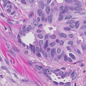
H&E tissue

#10167
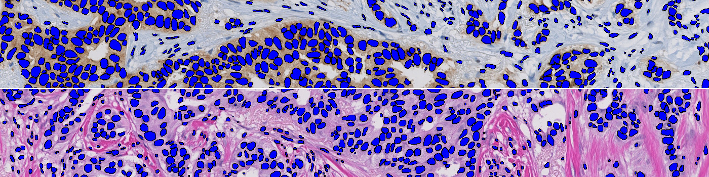
Developed for automatic segmentation of Nuclei in Brightfield images, exporting various shape measurements to assess morphology of each nucleus. Using deep learning (AI) for automatic nuclei detection in H&E and IHC stained tissue of various types including breast, prostate, liver, stomach and kidney, it is ready for use without additional training.
Users can customize the APP by adding other post-processing steps and/or output variables as needed.
H&E tissue
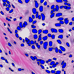
Nuclei segmentation in H&E
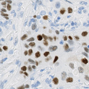
IHC stain (ER in breast cancer)
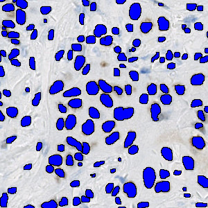
Nuclei segmentation
Quantitative Output variables
Exports:
Total number of nuclei
And for each Nucleus the output contains: