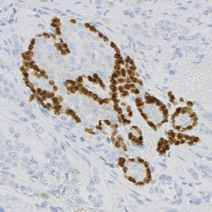
CK slide.

#10112
Whether a breast cancer is invasive or non-invasive will determine the treatment choices and how a patient might respond to the treatment he or she receives. Most breast cancers are invasive, but in some cases both invasive and non-invasive cancer can be seen in the same specimen, see [1]. In these cases, it is necessary to exclude non-invasive tumor components, such as ductal carcinoma in situ (DCIS), so that the biomarker expression can be accurately assessed within the invasive cancer.
The “Invasive Tumor Detection (VDS)” APP provide means for distinguishing between invasive tumor and non-invasive tumor based on two serial single stained slides with the myoepithelial cell nuclear marker p63 in one slide and a cytokeratin (CK) tumor marker, e.g. PCK, in another slide. With VirtualDoubleStaining™ the two slides are combined to include invasive tumor structures and exclude non-invasive tumor structures.
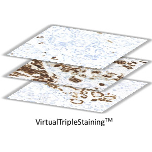
The “Invasive Tumor Detection (VDS)” APP can be combined with any biomarker APP using VirtualTripleStaining (VTS). With this approach biomarker analysis can be restricted to invasive tumor components.
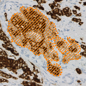
CK slide.
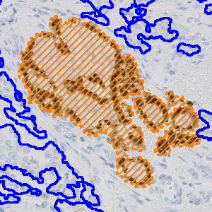
p63 slide.
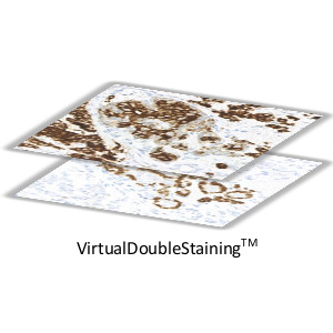
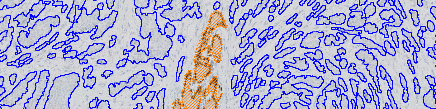
Using VirtualDoubleStaining™ the CK and p63 serial slides can be aligned, and a joint image can be created. This allows for the use of the information present on the individual images in a combined way.

p63 slide with overlay. Using the VDS principle non-invasive tumor structures, that are jointly positive for p63 and CK can be identified. This is step 1 of the APP.
Quantitative Output variables
The output variables obtained from this APP are:
Workflow
WITH VIRTUALDOUBLE STAINING™
Step 1: Align the serial sections stained with CK and p63, with CK on top
Step 2: Load and run the protocol “01 Non-invasive Tumor Detection” for detection of non-invasive tumor
Step 3: Load and run the protocol “02 Tumor Detection” for detection of invasive tumor
WITH VIRTUALTRIPEL STAINING™
Step 1: Align the serial sections stained with CK and p63 and the desired biomarker. The biomarker slide is on top with the CK slide as the second layer and the p63 as the third layer
Step 2: Load and run the APP “01 Non-invasive Tumor Detection” for detection of non-invasive tumor
Step 3: Load and run the APP “02 Tumor Detection” for detection of invasive tumor
Step 4: Load and run the APP suited for analysis of the slide stained with a desired marker within the invasive tumor components, e.g. APP “10004 – Ki-67, Breast Cancer”
Methods
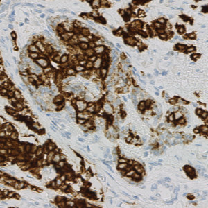
The APP consists of two protocols. The first protocol detects tumor structures positive for p63 and CK and classifies these as the non-invasive tumor structures (see FIGURE 4). The second protocol then detects all the remaining tumor present on the CK slides and classifies this as the invasive tumor components (see FIGURE 5-6).
If a biomarker slide, serial to the p63 and CK slides, is available, it is possible, using VirtualTripleStaining, to conduct an analysis where the information present on each of the slides is used in a combined way. For example, in the case of having a Ki-67 slide that is serial to the p63 and CK slides, it would be possible to identify the invasive and non-invasive tumor components on the Ki-67 slide based on the information present on the p63 and CK slides. With this method it would then be possible to do a Ki-67 quantification within the invasive tumor components only (see FIGURE 8).
Staining Protocol
The slides were stained with p63 (clone DAK-p63, brown chromogen) from Dako, PCK (clone AE1/AE3, brown chromogen) from Dako, and Ki-67 (clone MIB-1) from Dako.
Keywords
Invasive tumor detection, Ductal carcinoma in situ, VirtualDoubleStaining™, VDS™, VirtualTripleStaining, VTS, ER, PR, Ki-67, HER2, IHC
References
LITERATURE
1. Breastcancer.org. Non-Invasive or Invasive Breast Cancer. (Last modified: 19. September 2018).