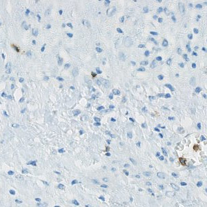
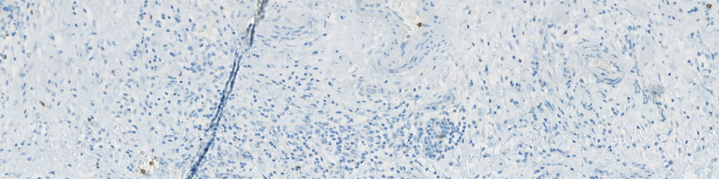
Selected view of ovarian CD57 stained tissue.
#10146
Natural Killer (NK) cells are a subtype of lymphocytes. They hold anticancer potential through tumor cell lysing and their prognostic role is being investigated in many cancers, with a perspective of a therapeutic potential. The prognostic role of tumor infiltrating NK cells in ovarian cancer, is being investigated at the Department of Clinical Pathology, Vejle Sygehus.
This APP can be used to evaluate the number or density of CD57 positive nuclei.
Selected view of ovarian CD57 stained tissue.
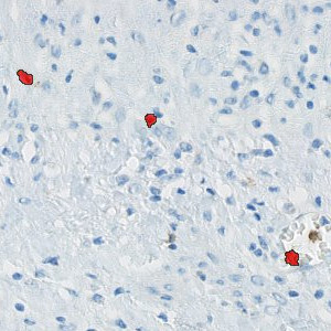
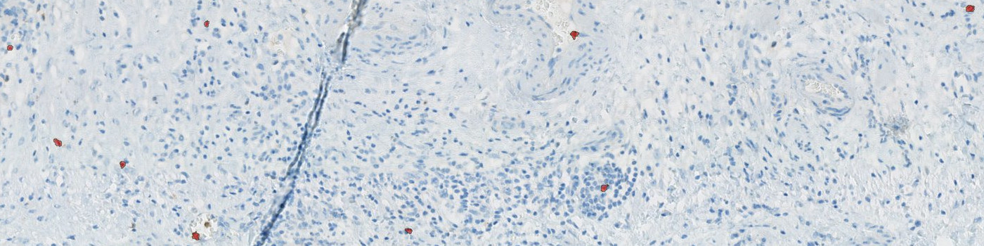
Final classification of NK cells (red). Brown structures with no blue core are discarded as CD57 positive cells.
Quantitative Output variables
The output variables obtained from this protocol are:
Workflow
Step 1: Manually outline regions of interest (ROIs)
Step 2: Load and run the APP for the quantification of CD57 nuclei
Methods
A NK positive cell is detected as a haematoxylin stained nucleus surrounded by DAB membrane stain. Initially, the protocol detects haematoxylin stained nuclei surrounded by brown stain using a poly blob filter on the H&E haematoxylin band and a median filter on the HDAB-DAB band. The DAB membrane stain is emphasized by a mean unsharp filter, thereby excluding blurry background stain. Then, positive nuclei are detected based on size, shape, and amount of surrounding DAB stain. Final NK cells are seen in FIGURE 2.
Staining Protocol
Thermo Fisher Scientific, Invitrogen (Roche Ventana Benchmark platform).
Keywords
CD57, NK cells, Natural Killer Cells, Ovarian Cancer, Cell Quantification, Image Analysis
References
USERS
This APP was developed for Dr. Jon Røikjær Henriksen, Department of Clinical Pathology, Vejle Sygehus.
LITERATURE
There are currently no references.