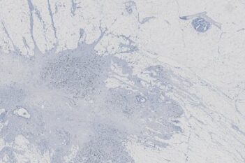
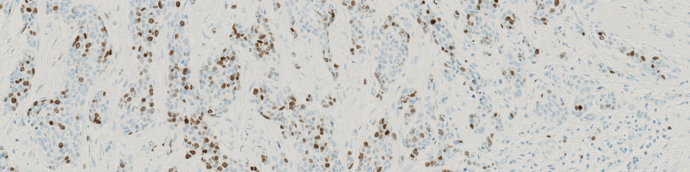
Invasive tissue detected with Ki-67 stain

#10162
This Quickstart APP will outline invasive tissue across a range of tissues in IHC-stained brightfield images. The Tumor Detection APP can be used after the Tissue Detection APP and prior to any of the Nuclei detection APPs. It has been tested with images of lung, breast and head and neck cancer and exports the total area of detected tumor in mm².
Invasive tissue detected with Ki-67 stain

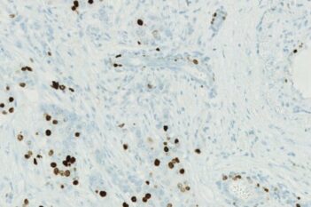
Invasive tissue detected in an ER-stained breast cancer slide

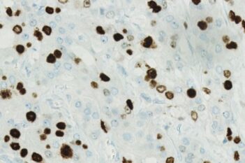
Highly positive slide with PR stain with detected tumor
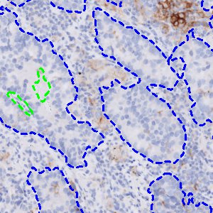
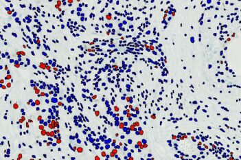
Lung tissue (PD-L1 negative) with segmented tumor regions
Quantitative Output variables
Total Tumor Area in mm²