Oncotopix® Discovery
Advancing scientific discovery
Oncotopix Discovery is designed for everyone and allows Researchers with tissue-based images to tackle both simple and complex datasets to generate reliable quantitative results.
Find out how easy it is
“Visiopharm not only saves time in analysis, it’s also more intuitive to use, it’s flexible and easier to train other users.”
Derick Vollmer, Manager of Digital Pathology at StageBio
The Oncotopix Discovery Workflow

“Another major advantage that influenced our decision was the exceptional support provided by Visiopharm. They respond quickly and efficiently to any queries or issues, which has been incredibly reassuring.”
Didier Merciris, Senior Project Manager at NovAliX

Where do you want to start?


Load images
Stay flexible
with the choice of your
imaging system
All major image formats:
Aperio (SVS, AFI)
Nikon (ND2)
3DHistech (MRXS)
Olympus / Evident (VSI)
Hamamatsu (NDPI, NDPIS)
Leica (SCN)
Zeiss (CZI)
Ventana (BIF)
Philips (iSyntax, i2Syntax)
JPG, TIF, BMP, OME.TIFF
And many more…
Import annotations
Utilize existing analysis pipelines and then continue where they leave off.
Transfer and use existing annotations from other platforms. Build classifiers by training on result masks.
Easy transition
of projects from
other analysis systems
“This highlighted one of the main benefits of using Visiopharm: which is the flexibility we have with APP design.”
Karen McClymont, Image Analysis Project Manager at OracleBio
Define analysis
Preconfigured APPs
Get started easily – Use our pretrained nuclear detection APPs or modify them to your needs using your expert knowledge. Add annotations or layers to tailor the APP to your specific needs.
Available for brightfield (H&E, IHC), immunofluorescence (IF, DAPI), imaging mass cytometry (IMC).
Intuitively explore even complex tissue data
Easy-to-use AI deep learning is standard in all of our research applications.
With Oncotopix Discovery, anyone with an understanding of tissue morphology can train an AI deep learning APP to get accurate and reproducible results.
All you need is your expert biological knowledge.
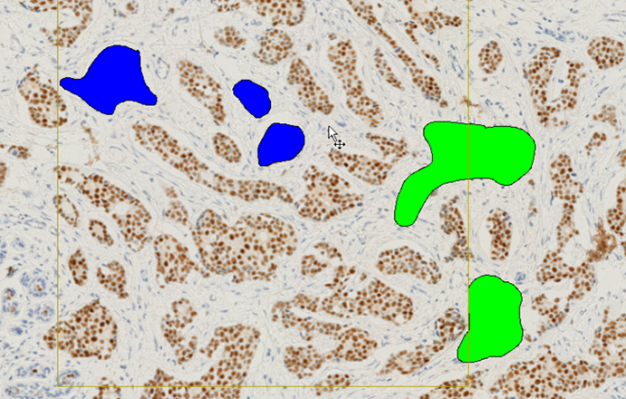
See it
Apply your tissue expertise and annotate the structures you want to identify. Our intuitive drawing tool allows for quick and seamless annotation.
No coding, no time-consuming training, no image analysis experience required.
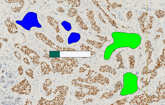
Train it
Leave the rest to Oncotopix Discovery.
The software will characterize the best features to robustly detect your structures even across heterogeneous tissues.
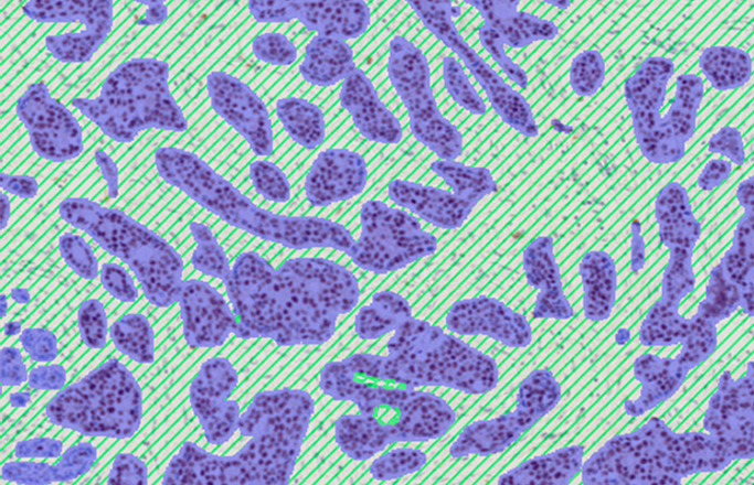
Find it
Once trained, the APP can be applied to large datasets, using batch processing for walk-away analysis.
Our deep learning APPs are designed to be enriched and reused. You can re-annotate regions and the APP will grow with your research.
Flexible analysis for tailored research
“Before we were always having to fit the study design to the analysis capabilities, whereas now we can fit the analysis approach to the requirements of the study.”
James Clay, Digital Pathology Manager at HistologiX
Analyze images
Batch mode
Run batch processing across entire studies with any number of research APPs.
Queue analysis tasks and batch process images in the background while simultaneously working on other tasks in parallel.
Parallel processing
Improve productivity with high speed processing of image analysis jobs. Oncotopix Discovery makes optimal use of the computational resources available where it is installed for parallelized, fast processing.
Explore results
Trust your results
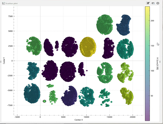
Review your cells and results in interactive plots and images simultaneously to QC your data analysis.
Clicking on a dot in a graph or plot will open the respective image or TMA and show the corresponding cell in the image context as well as a thumbnail of that cell in a gallery. Verify outliers and make sure that phenotypes are set correctly.
Dive into your data
Filter plots for specific regions, features or split view between additional treatment groups or tissue data.
Export data and images
Oncotopix Discovery exports all data, images and plots in standard formats for reports or publications.
Perform subsequent analysis in external data analysis pipelines through the .tsv files, which contain all detailed exports as saved to the database.
“With Visiopharm’s AI, we can now offer more in-depth and histologically relevant data, more structures, and functional regions of interest.”
Connor McCracken, Research Scientist at HistologiX
Additional modules
Tissuearray
Tissuearray is Visiopharm’s comprehensive automated solution for TMA analysis managing all metadata of the single cores for data correlation. It provides you with customizable grid layers for an easy and quick workflow. Import grid layouts to add metadata or import block designs from TMA spotters.
Tissuealign
Combine different sections or stain cycles into one flexible analysis.
Precise alignment of multiple serial sections of any image modality.
Align brightfield IHC or ISH with H&E and even IF or IMC images.
World-class service and support
Learn from our experts
Easy onboarding with our Academy courses or on-demand training session opportunities. Use our comprehensive training material and documentation for self learning and problem solving.
Continuous improvements through
regular software updates and maintenance with our Software Maintenance Service (SMS).