Stay curious

Whether you are working with drug discovery, drug development, spatial biology, or clinical research, our versatile tissue analysis software lets you build the exact analysis algorithms you need to unlock the secrets of your research.
Powerful deep learning – Easy to apply
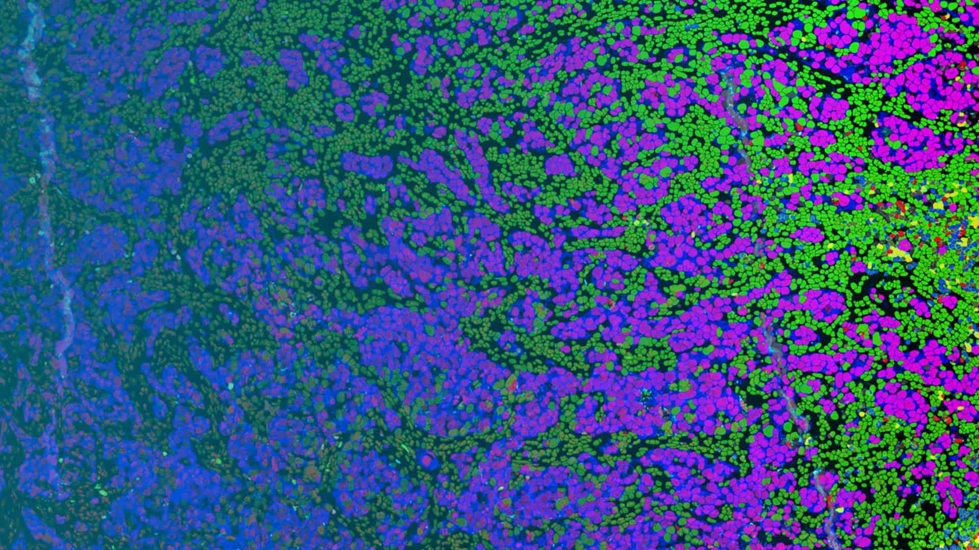
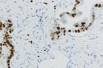
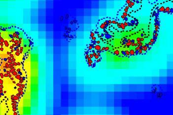
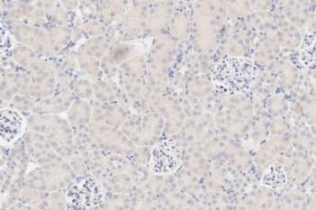
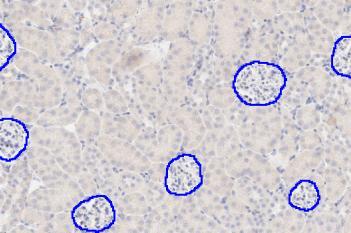
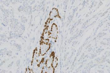
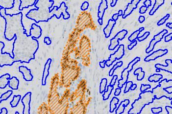
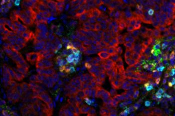
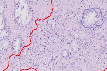
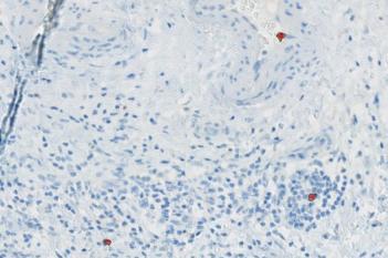
Integrating deep learning to image analysis allows robust and reliable detection of structures, which due to a high natural heterogeneity have been challenging to detect with machine learning only.
Deep learning based algorithms identify rules from the underlying patterns in the image, using examples instead of code.
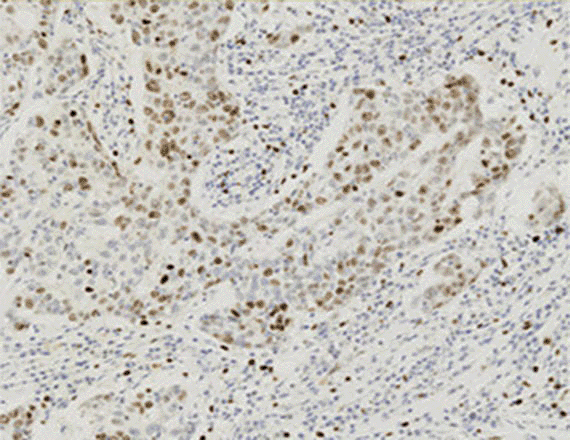
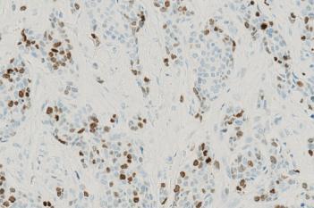
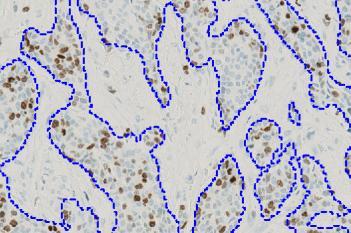
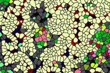
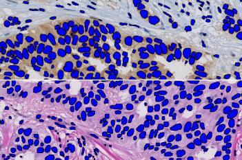
Our tissue image analysis platforms feature 14 preconfigured Quickstart APPs, which perform standard analysis steps like tissue detection and cell segmentation in just a few clicks.
These APPs are available for brightfield, fluorescence, IMC, and GeoMx® DSP, enabling you to dive right into the analysis.

“I chose Visiopharm because this software is different than all the others, it’s very versatile. It’s different than the black box software which comes with a pre-programmed set of modules.“
Nathan Su, Acepix Biosciences, USA
Explore our example APPs for Research
Get answers quickly with a wide array of APPs, amend existing APPs or create entirely new ones to fit your unique research questions.

Show all available APPs
Powerful data exploration tools
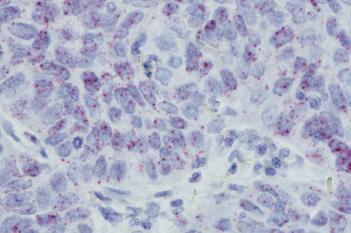
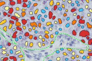
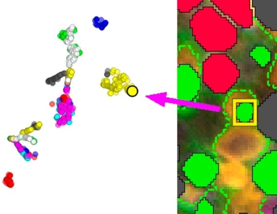
With Visiopharm software you get not only an ever-expanding suite of sophisticated analysis algorithms, you also get essential interactive data exploration tools to make sense of the results.Any selected data point in your plots will link back to the respective cell in the image to allow quick verification of the results. All plots can be adjusted to display each tissue compartment, phenotype, biomarker, or cell, as needed to properly interrogate results.
Aligning consecutive slides
The Tissuealign™ analysis module gives the ability to align and subsequently analyze digitized serial sections. This allows co-localization studies of multiple biomarkers through virtual multiplexing independently of image modality.
Align and co-analyze multiplex IF with H&E or IHC brightfield to get the most our of your data.
Speed that meets your needs
Run batch processing across entire studies with any number of research APPs. The software makes optimal use of the computational resources available where it is installed for parallelized, fast processing. Review all results interactively linked to the tissue sections or TMA core positions.
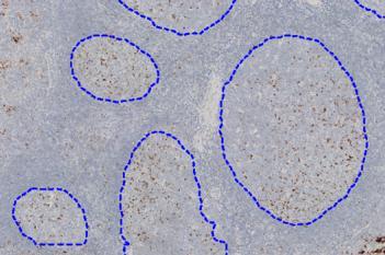
Tissue microarrays (TMA)
Tissuearray™ is one of the most comprehensive, dedicated solution available on the market. A guided workflow allows for managing cores and importing grid designs as well as apply any analysis solution in batch processing.
In combination with our IVDR diagnostic APPs, Tissuearray is validated for in vitro diagnostic use (CE IVD) in Europe. All other applications are for research use only.