Learn more about our powerful image analysis software for research pathology and diagnostics.
Software features
scroll down
Learn more about our powerful image analysis software for research pathology and diagnostics.
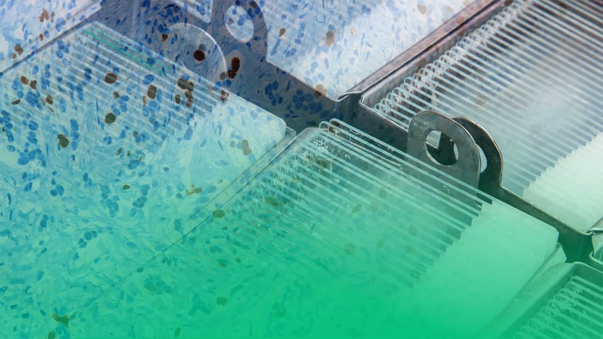
The Tissuealign™ analysis module gives the ability to align and subsequently analyze digitized serial sections. Tissue alignment is key for tumor cell detection and for tumor microenvironment studies.
Tissuearray™ is by far the most comprehensive, dedicated solution available on the market.
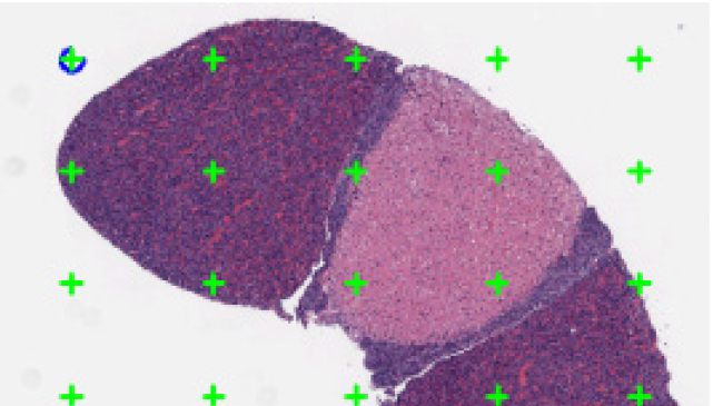
Easy, robust, and automated tumor-stroma separation.
The Visiopharm Viewer handles the most extensive support of whole slide formats and supports field of view (FOV) image types. The Viewer supports advanced image analysis for brightfield, fluorescence as well as multispectral image modalities.
The Visiopharm platform uses an “engine” developed with speed, scalability, and cost-efficiency in mind. The platform is available in different configurations so that diagnostic and research laboratories can match specific throughput requirements and grow in a logical and highly cost-efficient way.
![]()
Improve productivity in laboratories with high speed processing of image analysis jobs.
![]()
Queue analysis tasks and batch process images in the background while simultaneously working on other tasks in parallel.

Run batch processing across entire studies with any number of research APPs. The software makes optimal use of the computational resources available where it is installed for parallelized, fast processing. Review all results interactively linked to the tissue sections or TMA core positions.
Quantify structural information like volume, area, length and numbers from many types of tissue.
In addition to automatic tissue detection, utilize powerful tools for hot-spot detection, rare event detection, and more. With over 100 APPs developed, find the tools you need to analyze cells, regions and tissues across a wide range of applications.
The Visiopharm platform (Oncotopix) has become a national platform allowing use of semi-quantitative diagnostic IHC APPs in Denmark, with growing adoption across the globe. The platform is validated for Dako, Leica, and Ventana IHC/ISH reagent kits and all major slide scanner platforms. Plus, interfacing with a lab information system (LIS) is straightforward.
Visiopharm’s platform integrates seamlessly with a laboratory’s existing LIS/LIMS, IMS, PACS or VNA system; technologists and pathologists will access the solution through this familiar environment. Diagnostic pathology labs protect their LIS investment, and can now effectively launch digital Pathology into real life, diagnostic workflows.
All virtual slides associated with a case are automatically synchronized, linked for simultaneous viewing, and then presented in the pathologist workstation. Analysis results are presented as an overlay on the images for easy, transparent review.
Securely manage your data, share between experts, and search across experiments, studies, or institutes. Visiopharm software is integrated with many IMS/LIMS systems, allowing you to use cloud-based or on-premise management systems.