See it
Use your biological expertise to annotate objects of interest using our drawing tool. Background labels are generated automatically.
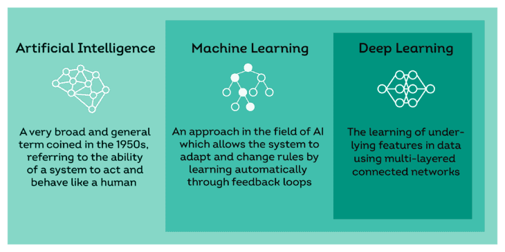
Deep learning is part of a broader family of artificial intelligence (AI) and machine learning methods. Deep learning is based on the latest technology and a much more advanced approach as it learns from underlying features in data using deep neural networks.
Oncotopix Discovery comes with pre-trained nuclear algorithms for brightfield and IF stained tissue sections – saving time and the staining of multiple sections. Quick start your specific analysis set up using those in three easy steps.
With convolutional neural networks (CNNs), a type of deep learning algorithms that are taught to recognize specific patterns, the underlying features and rules are learned directly from the images.
The deep learning technology in Visiopharm’s AI image analysis platform has been specifically developed for the field of histopathology, so you are able to apply, train and create high-quality deep learning algorithms to obtain breakthrough results in your field of work.
With our platform, you get the power of state-of-the-art deep learning to solve your problems – without having to write a single line of code.
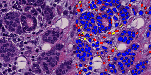
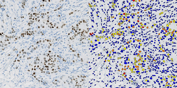
Our pre-trained nuclear detection modules facilitate automatic cell and nuclei identification on H&E and IHC stained tissue sections – saving time and staining of multiple sections.
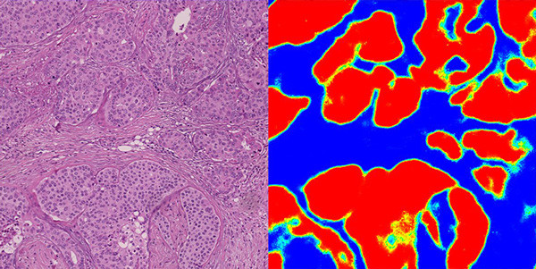
Obtain objective and reproducible results with our deep learning algorithms for any quantitative cell type analyses in combination with e.g. tumor and stroma segmentation.
Most cancer applications involve cell identification (classification + segmentation) and tissue compartmentalization. Manually solving this task is time-consuming, and highly challenging for the untrained eye, and often leads to subjective results. It is no longer the case with deep learning.
Develop image analysis protocols and workflows without the need for AI expertise. Use our unique authoring capabilities to tailor an existing APP to suit your needs, or to build your solution that delivers precise and accurate information without needing any programming experience.
The modular design of the software allows you to combine APPs and build a solution that answers your specific questions.
Visiopharm’s Oncotopix Discovery makes it a lot easier to solve challenging image analysis problems in tissue-based research. Using the Extended Author module, expert users can customize and control deep learning topologies such as training parameters, network architecture, and more.
This module gives access to the latest breakthroughs in AI and Deep Learning, representing the most comprehensive and configurable platform for artificial intelligence-based image analysis for digital pathology.
Understanding the biology of tissues calls for a complete toolbox of artificial intelligence techniques. Oncotopix Discovery is equipped with three types of deep learning classifiers. No matter which type of objects you need to classify, the full selection of AI tools is available at the click of a button.