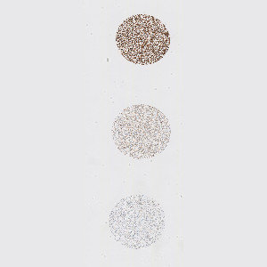
Raw image of cell line cores with different HEr2 expression.
#10105
The availability of genetically defined reference materials, offers an industry standard for development and quality control of IHC assays, directly, thereby improving the accuracy and reproducibility. Using engineered cell lines as quality control material to assess the performance of IHC assays eliminates the variability associated with patient-derived reference standards.
This APP can be used for quality control of HER2 negative (i.e. 0), 1+, 2+ and 3+ cell line material to ensure that each cell line block complies with the expected HER2 expression before the material is used as reference standard for IHC assays. The APP quantifies the HER2 expression in each cell line core and relates this expression to a known reference value for each cell line expression level.
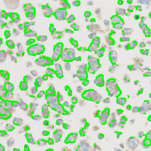
Raw image of cell line cores with different HEr2 expression.
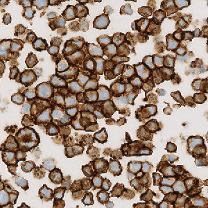
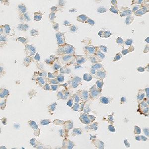
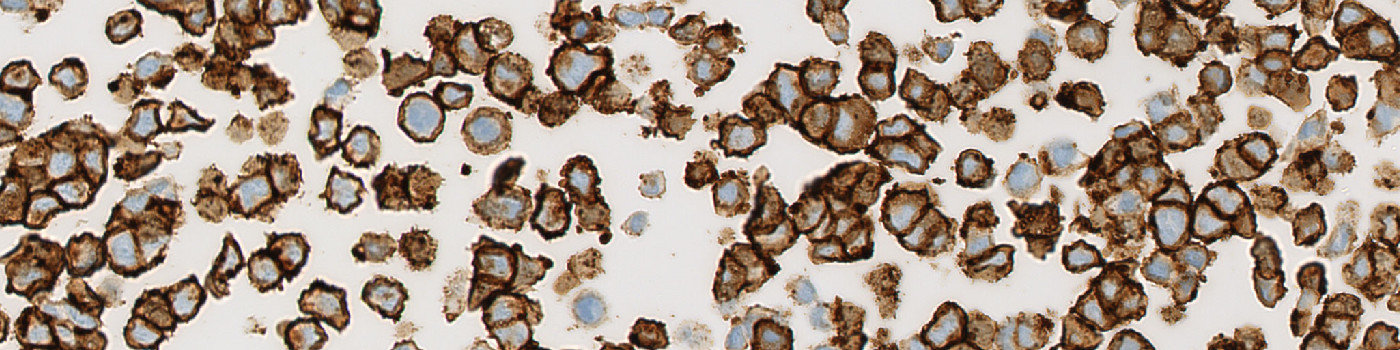
Cell line with IHC stained membranes, testing for HER2.
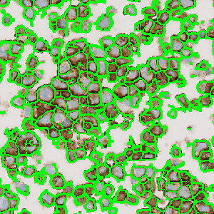
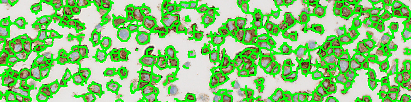
The image from FIGURE 2 analyzed with the APP. The cell line core expresses strong membrane staining and is categorized as a 3+ core.
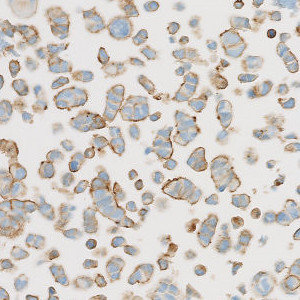
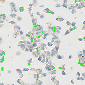
Cell line with IHC stained membranes, testing for HER2.
Quantitative Output variables
The output variables obtained from this protocol are:
Methods
First, a pre-processing step identifies the stained membrane pixels that contribute to linear structures in the image. Then, segmentation rules are employed identifying the membrane segments in the image. The segmentation is based on the intensity of brown in the pixels and the correct dimensions of linearity. After identifying pixels that constitute to brown linear structures and make up part of a membrane segment, post-processing steps are employed. The post-processing steps are used to skeletonize the membrane, removing small membrane fragments and merging membrane fragments which are not perfectly connected. From the membrane fragments remaining after post-processing, the connectivity can be calculated. Finally, the connectivity is translated into a classical HER2 score of 0, 1+, 2+ or 3+ based on known connectivity reference values for HER2 cell line cores expressing a score of either 0, 1+, 2+ or 3+.
Staining Protocol
There is no staining protocol available.
Keywords
Human Epidermal Growth Factor Receptor 2, HER2, cell line, quality control, reference standards, image analysis.
References
USERS
The APP was developed for Horizon cell lines.
LITERATURE
There are currently no references.