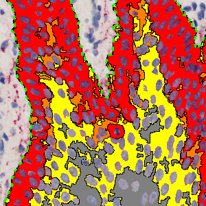
Part of epidermis stained with CK16
#10152
Cytokeratin-16 is a type I cytoskeletal protein. It is used as a marker for epidermal hyperplasia, a characteristic which is associated with various inflammatory skin diseases such as atopic dermatitis and psoriasis, see [1]. In contrast, CK16 protein expression in normal epidermis is very low or absent.
This protocol can be used for quantifying the epidermal expression of CK16. It divides the CK16 staining intensity into four categories: no staining (negative), low staining intensity (1+), medium staining intensity (2+), and high staining intensity (3+). An intensity score is calculated based on the categories.
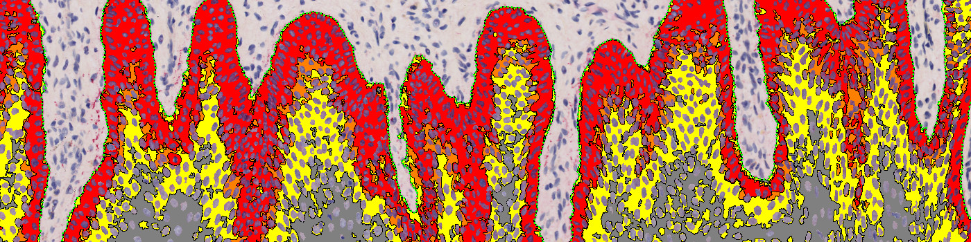
Part of epidermis stained with CK16
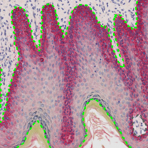
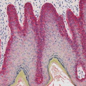
Manual outline of epidermis
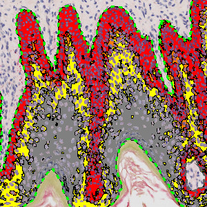
>CK16 staining intensity categorization within the epidermis with exclusion of nuclei
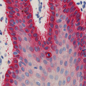
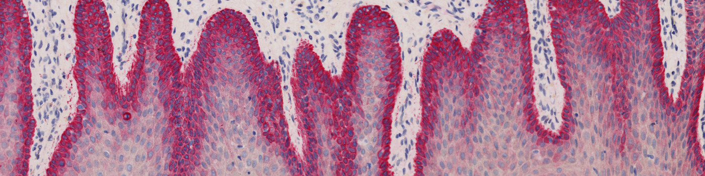
Microscopic view of epidermis stained with CK16
Quantitative Output variables
The output variables obtained from this protocol are:
Workflow
Step 1: Manually outline the epidermis
Step 2: Load and run the APP “10152 – CK16, Skin damage” for the quantification of CK16 staining intensity
Methods
The nuclei present in the epidermis are detected using a polynomial blob filter on a blue-enhancing feature image. The detection allows for exclusion of the nuclei before quantifying the CK16 staining intensity. A feature image, which revolves around red staining, is used for quantification of CK16 intensity. The intensity is divided into four groups ranging from negative (no staining) to strongly positive (3+) based on a threshold classifier and morphological post-processing steps is used for slight smoothing of the stain to handle grainy areas.
Staining Protocol
There is no staining protocol available.
Keywords
CK16, Skin, Skin damage, Dermatology, Digital pathology, Image analysis, IHC
References