Cellular and Metabolic Profiling the Tumor-Stroma Interface via High-Dimensional Spatial Proteomics and Image Analysis
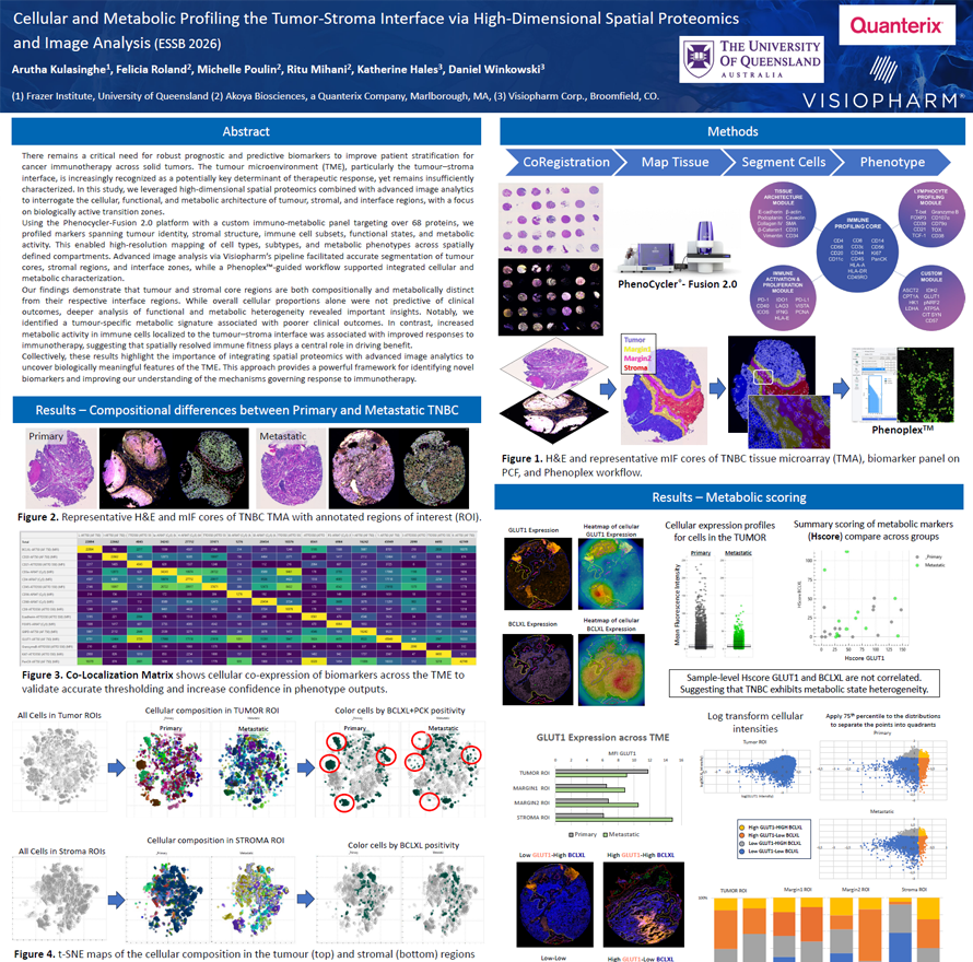
There remains a critical need for robust prognostic and predictive biomarkers to improve patient stratification for cancer
immunotherapy across solid tumors. The tumour microenvironment (TME), particularly the tumour–stroma interface, is increasingly recognized as a potentially key determinant of therapeutic response, yet remains insufficiently characterized. In this study, we leveraged high-dimensional spatial proteomics combined with advanced image analytics to interrogate the cellular, functional, and metabolic architecture of tumour, stromal, and interface regions, with a focus
on biologically active transition zones.
Using the Phenocycler-Fusion 2.0 platform with a custom immuno-metabolic panel targeting over 68 proteins, we profiled markers spanning tumour identity, stromal structure, immune cell subsets, functional states, and metabolic activity. This enabled high-resolution mapping of cell types, subtypes, and metabolic phenotypes across spatially defined compartments. Advanced image analysis via Visiopharm’s pipeline facilitated accurate segmentation of tumour
cores, stromal regions, and interface zones, while a Phenoplex -guided workflow supported integrated cellular and
metabolic characterization.
Our findings demonstrate that tumour and stromal core regions are both compositionally and metabolically distinct from their respective interface regions. While overall cellular proportions alone were not predictive of clinical outcomes, deeper analysis of functional and metabolic heterogeneity revealed important insights. Notably, we
identified a tumour-specific metabolic signature associated with poorer clinical outcomes. In contrast, increased metabolic activity in immune cells localized to the tumour–stroma interface was associated with improved responses to immunotherapy, suggesting that spatially resolved immune fitness plays a central role in driving benefit.
Collectively, these results highlight the importance of integrating spatial proteomics with advanced image analytics to uncover biologically meaningful features of the TME. This approach provides a powerful framework for identifying novel biomarkers and improving our understanding of the mechanisms governing response to immunotherapy.
Arutha Kulasinghe1, Felicia Roland2, Michelle Poulin2, Ritu Mihani2, Katherine Hales3, Daniel Winkowski3
- Frazer Institute, University of Queensland
- Akoya Biosciences, a Quanterix Company, Marlborough, MA
- Visiopharm Corp.,