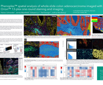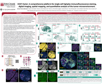

This white paper presents a detailed analysis using Phenoplex to study a highly multiplexed 30-biomarker assay of colorectal cancer (CRC) tissue, focusing on the spatial distribution of cytotoxic GZMB+ cells within CD8+ T cells and CD56+ NK cells. The study identifies these immune cells and examines their proximity to cytokeratin-expressing epithelial cancer cells. By segmenting the tissue into eight specific regions based on HLA-DR expression, the analysis reveals distinct patterns in immune cell infiltration and proximity, highlighting higher densities of cytotoxic T cells in HLA-DR- regions compared to HLA-DR+ regions. These findings suggest that the immune landscape and spatial relationships in the tumor microenvironment (TME) are crucial for understanding CRC pathology and potentially guiding therapeutic strategies.


