Advancing research through accurate
and reproducible tissue image analysis
powered by deep learning
Oncotopix® Discovery
Advancing research through accurate
and reproducible tissue image analysis
powered by deep learning
“Advances in deep learning […] have enabled the extraction of previously hidden information directly from routine histology images of cancer, providing potentially clinically useful information.”
Echle et al., DOI: 10.1038/s41416-020-01122-x
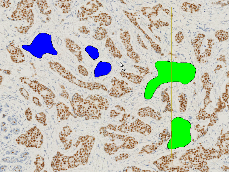
Apply your tissue expertise and annotate the structures you want to identify. Our intuitive drawing tool allows for quick and seamless annotation.
No coding, no time-consuming training, no image analysis experience required.
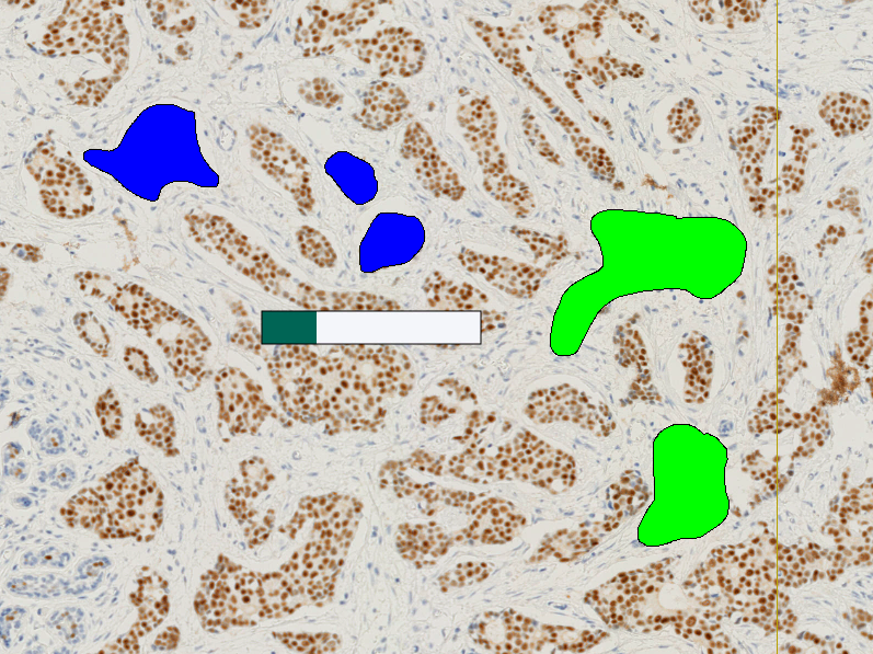
Leave the rest to Oncotopix Discovery. The software will characterize the best features to robustly detect your structures even across heterogeneous tissues.
Once trained, the APP can be applied to large datasets, using batch processing for walk-away analysis.
Our deep learning APPs are designed to be enriched and reused. You can re-annotate regions and the APP will grow with your research.

“The huge benefit of Visiopharm is that you can develop your own custom algorithms a lot more efficiently than you can with other software products. The possibilities are virtually endless.“
Stefan Hamann, PhD, Senior Principal Scientist at Boehringer Ingelheim Pharmaceuticals
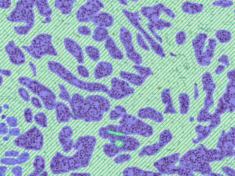
Pre-trained knowledge
Powerful pre-trained nuclei segmentation APPs, suitable for brightfield and fluorescence applications, can be further tuned for even more specificity.
Oncotopix Discovery is powered by Visiopharm’s best-in-class image analysis platform, combining 20 years of tissue expertise with innovative state-of-the-art technology. By combining your scientific knowledge with Visiopharm’s analysis expertise, you can generate the reliable quantitative data needed for your research studies and publications.
Tissue Microarrays (TMAs) are an effective method for high-throughput tissue analysis. Tissuearray is Visiopharm’s comprehensive automated solution for TMA analysis managing all metadata of the single cores for data correlation. Tissuearray reads block designs from professional TMA spotters, links IDs with individual TMA cores and automatically aligns all cores in linked sections based on information from the block design.
Our 20 years of tissue experience, patented technology and the latest applications of AI deep learning have empowered thousands of projects, yielding results that produced hundreds of publications and countless scientific breakthroughs.
Learn more