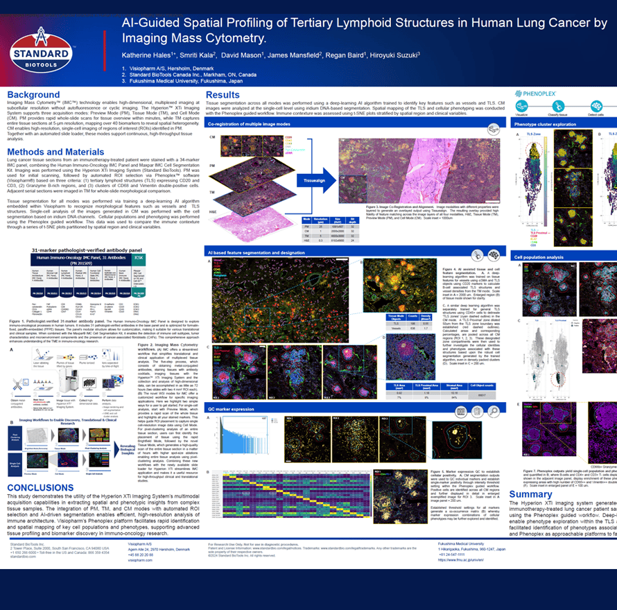
Imaging Mass CytometryTM (IMCTM) technology enables high-dimensional, multiplexed imaging at subcellular resolution without autofluorescence or cyclic imaging. The HyperionTM XTi Imaging System supports three acquisition modes: Preview Mode (PM), Tissue Mode (TM), and Cell Mode (CM). PM provides rapid whole-slide scans for tissue overview within minutes, while TM captures entire tissue sections at 5-μm resolution, mapping over 40 biomarkers to reveal spatial heterogeneity. CM enables high-resolution, single-cell imaging of regions of interest (ROIs) identified in PM. Together with an automated slide loader, these modes support continuous, high-throughput tissue analysis.
Katherine Hales1+, Smriti Kala2, David Mason1, James Mansfield2, Regan Baird1, Hiroyuki Suzuki3
- Visiopharm A/S, Hørsholm, Denmark
- Standard BioTools Canada Inc., Markham, ON, Canada
- Fukushima Medical University, Fukushima, Japan