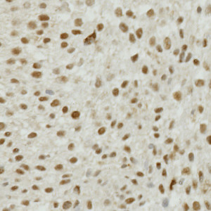
Field of view showing a tumor region in an image of a bladder tissue TMA core stained by IHC for Survivin.
#10014
Survivin is a multifunctional protein. It has been shown to inhibit apoptosis, to regulate cell division, and to enhance angiogenesis, see [1]. Studies have shown the expression of Survivin in non-muscle invasive bladder cancer to associate with progression to muscle-invasive disease, see [2]–[5]. Survivin has also been investigated in several other cancers. It has been discussed whether total, nuclear, or cytoplasmic Survivin expression should be evaluated in relation to clinical outcome. When using this protocol, we focus on the nuclear expression of Survivin.
This APP can be used for quantifying the nuclear expression of Survivin. The approach to calculating expression used here, was demonstrated to have a statistically significant correlation to manual scoring in a study involving 300 patients (publication pending).
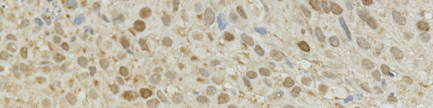
Field of view showing a tumor region in an image of a bladder tissue TMA core stained by IHC for Survivin.
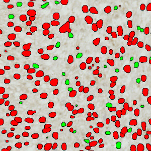
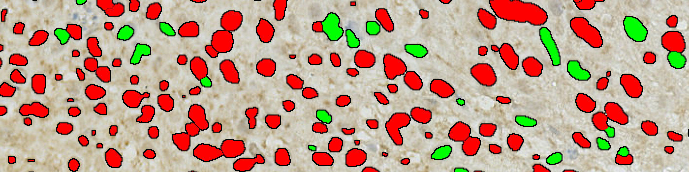
The same field of view as in Figure 1 after automatic segmentation for Survivin-positive (red label) and -negative (green label) nuclei.
Quantitative Output variables
The output variables obtained from this protocol include:
Methods
The method used for computing the Survivin expression starts by identifying nuclei using a novel pattern recognition method adapted from Kårsnäs et al see [6], which is followed by a step that separates adjacent nuclei, see [7].
The identified nuclei are classified as either positive or negative based on a computation of DAB intensity, obtained using color deconvolution. This is followed by a calculation of the positive area fraction as the area of positive nuclei divided by the total area of nuclei (within the outlined ROI). The average DAB intensity across all positive nuclei is measured and subtracted from 255, to have an association between large numbers and a high staining intensity, which is more intuitive. The final expression is calculated by multiplying the intensity with the area fraction.
The actual implementation of the pattern recognition allows the user to choose settings for nuclear detection sensitivity and classification into positive and negative, in order to account for differences in local staining protocols.
Optimal settings were determined for the specific tissue preparation protocol, and all tissue sections in the study were batch-processed with those settings.
It should be noted that this approach to quantifying biomarker expression is vulnerable to variations in protocols for tissue preparation (fixation time, tissue thickness, reagents, auto-stainer type and settings, etc.). Using this APP for TMA study designs, however, is robust for the cores within the TMA.
This APP requires manual outlining of tumor regions.
Additional information
This APP was developed for, and validated by, Niels Fristrup MD and Dr. Lars Dyrskjøt Andersen, Center for Molecular Clinical Cancer Research Department of Molecular Medicine (MOMA) Aarhus University Hospital.
Keywords
Survivin, bladder cancer, metastasis, immunohistochemistry, image analysis, quantitative, digital pathology
References
USERS
This APP was developed for, and validated by, Niels Fristrup MD and Dr. Lars Dyrskjøt Andersen, Center for Molecular Clinical Cancer Research Department of Molecular Medicine (MOMA) Aarhus University Hospital
LITERATURE
1. Duffy, M.J., et. al. Survivin: a promising tumor biomarker, Cancer Lett 2007, 249 (1), 49-60, DOI
2. Yin, W., et. al. Survivin nuclear labeling index: a superior biomarker in superficial urothelial carcinoma of human urinary bladder, Mod Pathol 2006, 19 (11), 1487-97, DOI
3. Ku, J.H., et. al. Expression of survivin, a novel inhibitor of apoptosis, in superficial transitional cell carcinoma of the bladder, J Urol 2004, 171 (2), 631-5, DOI
4. Karam, J.A., et. al. Survivin expression in patients with non-muscle-invasive urothelial cell carcinoma of the bladder, Urology 2007, 70 (3), 482-6, DOI
5. Fristrup, N., et. al. Cathepsin E, Maspin, Plk1, and Survivin are promising prognostic protein markers for progression in non-muscle invasive bladder cancer, Am J Pathology 2012, 180 (5), 1824-34, DOI
6. Kårsnäs, A., et. al. Learning histopathological patterns, Journal of Pathology Informatics 2011, 2 (2), S12, DOI
7. Jung, C., et. al. Segmenting Clustered Nuclei Using H-minima Transform-Based Marker Extraction and Contour Parameterization, IEEE Transactions on Biomedical Engineering 2010, 57 (10), 2600-4, DOI