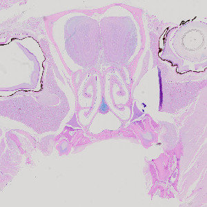
Cross section of rodent head, stained with AB/PAS.

#10115
Nasal mucous cell metaplasia (MCM) is a remodeling process where nasal respiratory epithelial cells undergo metaplasia to mucous cells. This causes an increased production of mucous, which leads to increased airway obstruction. Mucous cell metaplasia is often observed as a feature of allergic airways disease after exposure to an allergen. The mucosubstances in the respiratory epithelium are identified by staining with Alcian Blue (pH 2.5)/Periodic Acid-Schiff (AB/PAS or AB-PAS). Using this APP the degree of mucous cell metaplasia is quantified in the nasal respiratory epithelium as the volume of mucosubstances per length of basal lamina.
Cross section of rodent head, stained with AB/PAS.
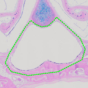
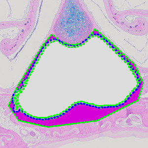
Close-up on nasal cavity with manually outlined region of interest.
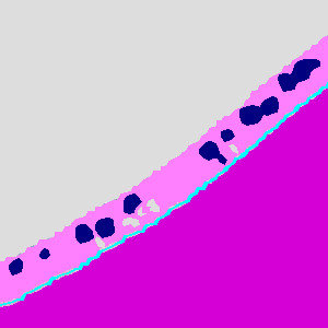
Results of analysis with the APP: “01 Tissue Detect”. The background, epithelial tissue and the remaining tissue are outlined.
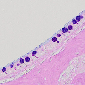
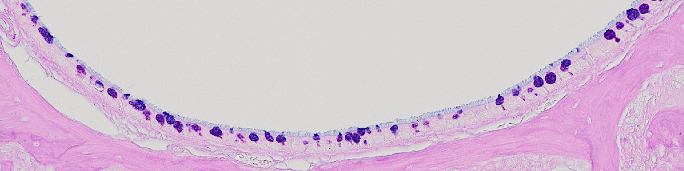
Close up of nasal epithelium.
Auxiliary APPs
APP: “01 Tissue detect”
APP: “03 Analyze”
Quantitative Output variables
The output variables obtained from this protocol are:
Workflow
Step 1: Manually outline the nasal cavity as the region of interest, see FIGURE 2.
Step 2: Load the APP for tissue detection “01 Tissue Detect” which outlines the background, epithelial tissue and remaining tissue.
Step 3: Load the APP for mucous detection “02 Mucous Detect” which identifies the basal lamina and mucosubstances inside the epithelial tissue.
Step 4: Load the quantification protocol “03 Analyze” which quantifies the relevant output parameters. Click the save button to transfer the results to the database.
Methods
The first image processing step takes place inside the manually outlined general region of interest (ROI), and involves the segmentation of the tissue into epithelial tissue and remaining tissue as shown in FIGURE 3. Secondly, the basal lamina is identified as the border between the epithelial tissue and the remaining tissue (see FIGURE 5). Afterwards the intraepithelial mucosubstances are identified based on a linear Bayesian classification combined with prior knowledge of the size of mucusubstances (see FIGURE 5). Finally, relevant output parameters such as the basal lamina length and the mucous area are quantified.
Staining Protocol
The staining protocol has not been specified.
Keywords
Mucous cell metaplasia, nose, nasal, respiratory, AB/PAS, AB-PAS, airway disease, quantitative, image analysis.
References
LITERATURE
There are currently no references.