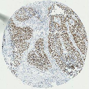
Overview of a single MCM7 stained core.
#10031
The DNA replication licensing factor MCM7 is part of the MCM-complex which ensures that DNA undergoes a single round of replication per cell cycle, see [3]. It has also been shown to unwind double-stranded DNA at the replication forks, see [4]. The MCM7 gene has been shown to be up-regulated in a number of cancers compared to normal tissue. Of special interest is that the gene has been shown to be overexpressed in prostate cancer compared with the normal prostate, and this overexpression has been associated with prostate cancer progression, see [5]. This relation of overexpression and progression has also been shown for bladder cancer, see [6]. SiRNA mediated knockdown of the MCM7 gene in bladder cancer cell lines has been shown to diminish proliferation, see [7].
This APP can be used for quantifying the nuclear expression of MCM7. The approach used here to calculate expression, have in a recent study by Fristrup et al, see [8] been shown to be highly prognostic in both a test cohort (n = 283) and a validation cohort (n = 576) of non-muscle invasive bladder cancer with long-term follow-up. The MCM7 APP was in the study shown to prognosticate patients very similar to manual scoring of the protein.
Overview of a single MCM7 stained core.
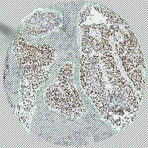
Same core as in FIGURE 1. The core has been automatically outlined, followed by manual editing to exclude stromal tissue.
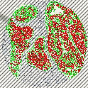
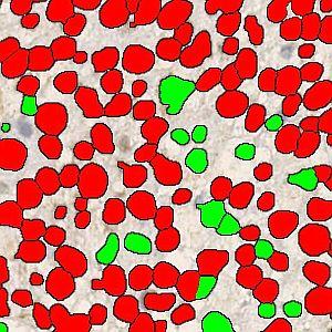
Same core as in FIGURE 1 and 2. The outlined area has been analyzed, where red denotes positive nuclei and green negative nuclei.
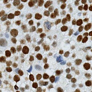
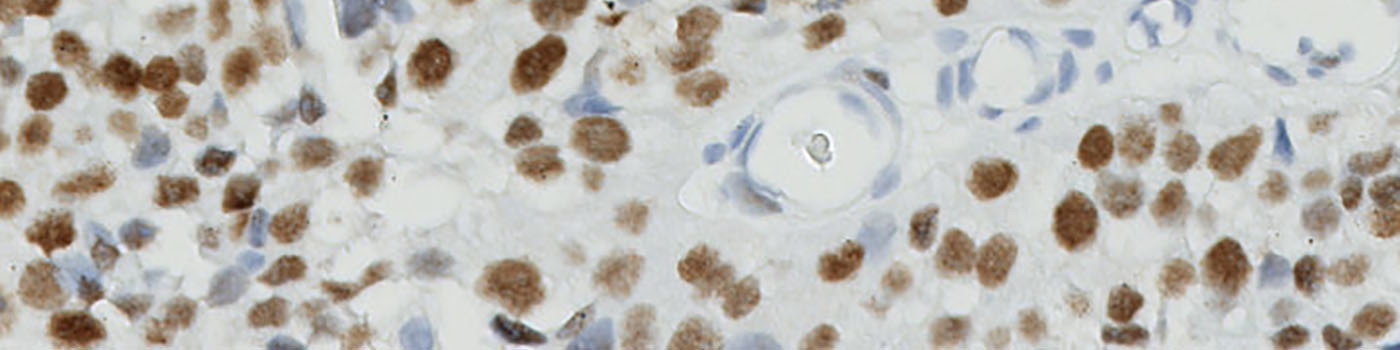
One field of view of the original image at 20X (scaled down to fit this space).
Quantitative Output variables
The output variables obtained from this protocol include:
Methods
The method used for computing the MCM7 expression starts by identifying nuclei using a novel pattern recognition method adapted from Kårsnäs et al see [1], which is followed by a step that separates adjacent nuclei, see [2].
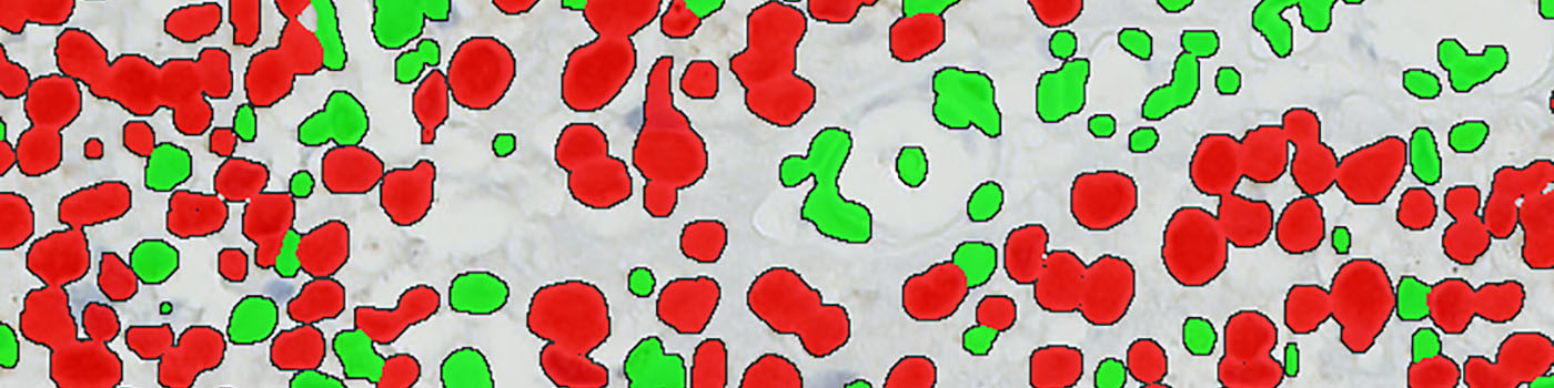
The identified nuclei are classified as either positive or negative based on a computation of DAB intensity, obtained using color deconvolution. This is followed by a calculation of the positive area fraction as the area of positive nuclei divided by the total area of nuclei (within the outlined ROI). The average DAB intensity across all positive nuclei is measured and subtracted from 255, to have an association between large numbers and a high staining intensity, which is more intuitive. The final expression is calculated by multiplying the intensity with the area fraction.
The actual implementation of the pattern recognition allows the user to choose settings for nuclear detection sensitivity and classification into positive and negative, in order to account for differences in local staining protocols. Optimal settings were determined for the specific tissue preparation protocol, and all tissue sections in the study were batch-processed with those settings.
It should be noted that this approach to quantifying biomarker expression is vulnerable to variations in protocols for tissue preparation (fixation time, tissue thickness, reagents, auto-stainer type and settings, etc.). Using this APP for TMA study designs, however, is robust for the cores within the TMA.
This APP requires manual outlining of tumor regions.
Additional information
This APP was developed for, and validated by, Niels Fristrup MD and Dr. Lars Dyrskjøt Andersen, Center for Molecular Clinical Cancer Research Department of Molecular Medicine (MOMA) Aarhus University Hospital.
Keywords
MCM7, bladder cancer, metastasis, immunohistochemistry, image analysis
References
USERS
This APP was developed for, and validated by, Niels Fristrup MD and Dr. Lars Dyrskjøt Andersen, Center for Molecular Clinical Cancer Research Department of Molecular Medicine (MOMA) Aarhus University Hospital.
LITERATURE
1. Dahl, A.L., et. al., Learning Dictionaries of Discriminative Image Patches, British Machine Vision Conference 2011, DOI
2. Jung, C., et. al., Segmenting Clustered Nuclei Using H-minima Transform-Based Marker Extraction and Contour Parameterization, IEEE Transactions on Biomedical Engineering 2010, 57 (10), 2600-4, DOI
3. Chong, J.P., et. al., Purification of an MCM-containing complex as a component of the DNA replication licensing system, Nature 1995, 375 (6530), 418-21, DOI
4. Ishimi, Y. A, DNA helicase activity is associated with an MCM4, J Biol. Chem. 1997, 272 (39), 24508-13, DOI
5. Ren, B., et. al., MCM7 amplification and overexpression are associated with prostate cancer progression, Oncogene 2006, 25 (7), 1090-8, DOI
6. Dyrskjøt, L., et. al., Gene expression signatures predict outcome in non-muscle-invasive bladder carcinoma: a multicenter validation study, Clin. Cancer Res. 2007, 13 (12), 3545-51, DOI
7. Toyokawa, G., et. al., Minichromosome Maintenance Protein 7 is a potential therapeutic target in human cancer and a novel prognostic marker of non-small cell lung cancer, Mol. Cancer 2011, 10 (1), 65, DOI
8. Fristrup, N., et. al., Multicenter validation of Cyclin D1, MCM7, TRIM29, and UBE2C as prognostic protein markers in non-muscle invasive bladder cancer (in review), American Journal of Pathology 2013, 182 (2), 339-49, DOI