Immune checkpoint inhibitors (ICIs) Pembrolizumab and Nivolumab have shown promising results in patients with recurrent or metastatic (R/M) head and neck squamous cell carcinoma (HNSCC). Nonetheless, only up to 20% of patients may benefit from them. Next generation predictive biomarkers of response to ICI therapies are currently needed.
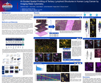
This retrospective study profiled pre-treatment tissue samples collected from R/M HNSCC patients (n=30) treated with Pembrolizumab or Nivolumab. Targeted spatial proteomics profiling was performed using NanoString’s GeoMx® Digital Spatial Profiler (GeoMx). To develop an informed region-of-interest (ROI) selection strategy for the GeoMx, we analyzed H&E images from serial sections and performed Oncotopix® Discovery software, which considered pathology inputs and machine learning to analyze H&E imagery and 4-channel GeoMx visualization inputs. We profiled 80 proteins simultaneously using the GeoMx across modules in cell death, immune activation, immune cell typing, immuno-oncology drug target, immune profiling, PI3K/AKT signaling, and pan-tumor markers. Next, we performed differential expression (DE) analysis of the proteins localized in the tumor and stromal compartments against response to therapy parameters of complete, partial, stable and progressive disease (according to RECIST criteria).
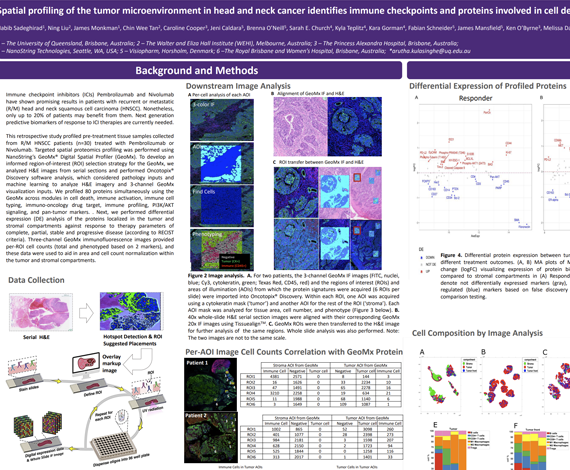
Four-channel GeoMx immunofluorescence images provided per-ROI cell counts (total and phenotyped based on 3 markers), and these data were used to aid in area and cell count normalization within the tumor and stromal compartments. We found that patients responsive to immunotherapy had higher expression levels of PD-L1, Bcl-2, BCLX, and BIM in the tumor, whereas VISTA, FOXP3, and CD66b were downregulated. In the stromal compartment, it was found that responders had higher expression levels of B7-H3, CD40, and SMA, but lower expression of PARP, S100B, and NY-ESO-1 in comparison to non-responders. In terms of best response analysis, patients with PR (n=5) had higher levels of PD-L1, ER-alpha, and CD68 expression, but lower levels of VISTA, CD27, and CD95/Fas in tumor regions than patients with PD (n=8). In the stroma, PD-L1, CD68, and HLA-DR were upregulated, while VISTA, BIM, and BAD were downregulated in patients with PR versus those with PD.
We found that informed ROI selection strategy aided in defining key features in the tumor microenvironment for comparative analysis across samples, and for data normalization methods. Moreover, we found that immune checkpoints and proteins involved in cell death signaling play important roles in the immune responsive tumor microenvironment of HNSCC based on response to immunotherapy.
Habib Sadeghirad1, Ning Liu2, James Monkman1, Chin Wee Tan2, Caroline Cooper3, Jeni Caldera4, Sarah Church5, Fabian Schneider4, James Robert Mansfield4, Ken O’Byrne3, Melissa Davis2, Brett Hughes6, Arutha Kulasinghe1
- The University of Queensland, Brisbane, Australia
- The Walter and Eliza Hall Institute, Melbourne, Australia
- The Princess Alexandra Hospital, Brisbane, Australia
- Visiopharm, Horsholm, Denmark
- Nanostring, Seattle, WA
- The Royal Brisbane and Women’s Hospital, Brisbane, Australia