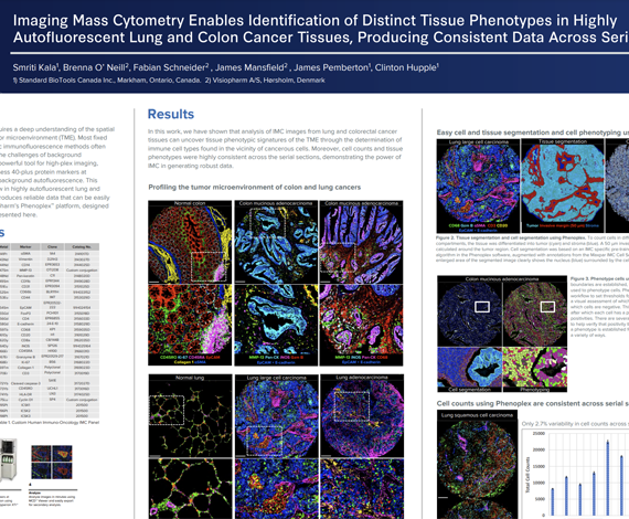
Successful implementation of immunotherapy requires a deep understanding of the spatial interactions between various cell types in the tumor microenvironment (TME). Most fixed tissues are autofluorescent and staining with cyclic immunofluorescence methods often produces data that are difficult to analyze due to the challenges of background subtraction. Imaging Mass Cytometry™ (IMC™) is a powerful tool for high-plex imaging, utilizing CyTOF® technology to simultaneously assess 40-plus protein markers at subcellular resolution without spectral overlap or background autofluorescence. This study demonstrates a tissue phenotyping workflow in highly autofluorescent lung and colon cancer tissues using high-plex IMC, which produces reliable data that can be easily analyzed.
Smriti Kala1, Brenna O’ Neill2, James Pemberton1, James Mansfield2, Fabian Schneider2, Clinton Hupple1
- Standard BioTools, Markham, ON, Canada
- Visiopharm, Hørsholm, Denmark