Transcript
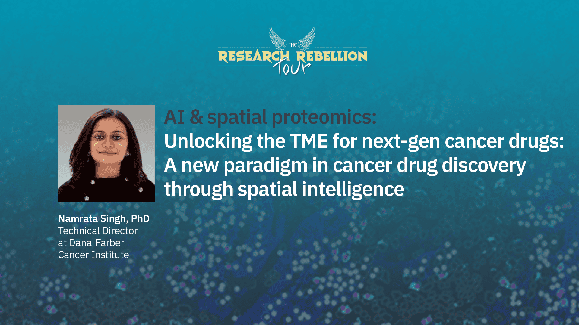
Welcome everybody. I’m Bettina Winkler from Visiopharm and I would like to welcome you to a virtual research rebellion tour. Today, we’re kicking off the series with doctor Namrata Singh, is Technical Director of the Dana Farber Cancer Institute Core Pathology Lab and also holds a concurrent research position at Harvard Medical School. She has nearly three decades of experience spanning cancer research, spatial biology and histological analysis, bridging the gap between technology and translational research.
Namrata will talk about how AI and spatial proteomics enable highplex protein level mapping of cells with intact tissue architecture providing insights into both expression and localization patterns, unlocking the tissue microenvironment for next gen cancer drugs. She will answer your questions after the talk, so please add any questions to the chat. With that, let’s get started.
Hello, everyone. My name is Namrata Singh. I’m a technical director at Dana Farber Cancer Institute at Pathology Core Lab.
It is a privilege to discuss the convergence of two or more transformative frontiers in modern oncology.
For decades, our understanding of tumor microenvironment has been limited to viewing it as a disorganized collection of cells. However, we now recognize that in the context of oncology and therapeutic resistance, location is paramount.
Much as an architect must grasp the intricate structure integrity of a building to refine its design, we must decipher the complex architecture of tumor to effectively dismantle it.
Today, I’m happy to present how we are leveraging artificial intelligence to bridge this gap, transforming raw data into spatial intelligence to catalyze the discovery of next generation cancer therapeutics.
The topics I’ll be covering in my talk today are tumor microenvironment and spatial protomics, why spatial protomics and AI matters for oncology.
I’ll be sharing some case studies and the workflows for drug target ID and biomarker discovery and some challenges associated with these methods.
To truly unlock the complexity of tumor microenvironment, we have developed a seamless end to end workflow that integrates highplex imaging with advanced computational analysis.
It begins with spatial omics phase where we utilize highplex immunofluorescence to capture the precise location of dozens of biomarkers simultaneously within a single tissue section.
This raw spatial data is then processed through our AI powered analysis engine, which performs automated cell segmentation, phenotyping, and neighborhood analysis to decode the social network of the tumor.
Finally, this leads us to actionable insights where we translate these spatial patterns into quantifiable data that can predict drug response, identify new therapeutic targets, and ultimately accelerates the delivery of next generation cancer treatment.
Tumor microenvironment.
Why do we need to know about tumor microenvironment?
Cancer is currently second most common cause of death in United States and is likely to become the most common in the future.
Tumor microenvironment is an ecosystem of immune cells, fibroblasts, vasculature, signaling molecules, and extracellular matrix that surrounds and interacts with tumor cells.
It actively shapes tumor growth, immune evasions, metastasis, and therapy resistance, making it a critical determinant of cancer progression.
Because of this, the TME is both a major barrier and a promising target for effective cancer therapies.
Spatial proteomics technologies have rapidly advanced and now key to Atlas scale projects enabling detailed mapping of tissue proteomes and enhancing our understanding of health and disease.
Spatial chromatomics encompasses a diverse array of technologies that vary in scale, resolution, sensitivity, offering researchers and clinicians a broad range of tools to investigate the spatially resolved proteome. The depth and resolution of spatial proteomics and in particular deep visual proteomics makes these technologies incredibly valuable in biomedical research.
Spatial biology is revolutionizing biomedical research by emphasizing three d relationships and arrangements of biological molecules within a tissue, exploring cell phenotypes, correlated locations, and cell cell interactions in the tissue microenvironment can help understand biological processes and disease pathogenesis.
Integration of technology and biology, why now?
Availability of high dimensional data set including multi omics, high performance computing, decline in the price of massively paralyzed computing power, innovative machine and deep learning algorithms.
These tools with biological data, researchers can better understand disease, identify new drug targets, and personalize medical treatments.
This convergence is accelerating discoveries and enabling more precise and predictive biology.
Now I’ll be sharing some case studies that I worked in my lab with you all.
I have nothing to disclose with respect to this presentation.
This slide illustrates the evolution of pathology at DFCI Core Pathology Lab across three generations.
The first generation relied on traditional glass slide diagnosis where pathologists manually examined samples under microscopes.
The second generation introduced whole slide imaging, allowing slides to be digitized and viewed on computers, improving storage and sharing of data. Third generation incorporated Phenocycler Fusion, a COIA platform, an AI based algorithm, Visiopharm. This AI based algorithms in pathology has assisted in analyzing digital images and identifying disease patterns.
Overall, this progression shows how technology is transforming pathology from manual analysis to intelligent data driven diagnostics in our core lab.
This slide shows six plex immunomarker panel on human tonsil tissue that I have worked on phenocycle fusion.
As we all know, tonsil tissue serves as an ideal model for validating multiplex spatial imaging because of its well organized tumor microenvironments, including B cell follicles, germinal centers, and T cell zones.
Using the cyclical imaging workflow of the phenocycler fusion platform, these six markers can be visualized within the same tissue section, enabling high resolution spatial mapping of proliferating immune populations and their organizations within the tonsillar architecture.
This multiplex approach highlights how spatial biology technologies can reveal cellular distribution, activation states, and interaction in complex lymphoid tissues.
Why Visiopharm? Visiopharm is a flexible platform. It allows development of custom applications, or you can also use a pre built solutions that are already provided with the software.
It reduces reliance on time consuming manual annotations required by other software.
It is very user friendly. No coding is required.
This makes it accessible to a wide range of users.
It enhances the reliability and consistency of data.
Last but not least, they have excellent customer support. They’re very responsive and they’re very effective in assisting you.
I use the co occurrence matrix workflow in Visiopharm to analyze spatial relationship between different phenotyped cell populations in multiplex tissue images. After performing cell segmentation and phenotyping, the workflow calculates pairwise proximity between cell types within a defined radius.
This generates a co occurrence matrix that highlights enrichment or avoidance of specific cell cell interactions.
This analysis also helps characterize cellular neighborhoods and spatial organization of tumor microenvironment. Case study one, glioblastoma.
An immunophenotyping panel was applied to give glioblastoma tissue using phenocyclofusion platform to characterize the spatial immune landscape of tumor microenvironment.
The panel includes c d four, c d eight to identify helper and cytotoxic T cell population, c d twenty for B cells, FOX p three for regulatory T cells, and HLA to access antigen presentation activity.
PANC K was incorporated to delineate epithelial tumor cell regions and provide structural context for immune cell localization.
This multiplex spatial profiling enabled high resolution mapping of immune cell distribution and their interactions within glioblastoma tissue. The resulting spatial data was analyzed using Phenoplex workflow to perform co occurrence and neighborhood analysis, enabling quantitative assessment of spatial relationship and cellular interaction within immune and tumor compartments of glioblastoma tissue.
Localization of CD four and CD eight T cells was egg assessed relative to pan c k tumor regions to evaluate immune cell distribution within and around tumor compartments.
This spatial mapping enabled the identification of T cell infiltration patterns in relation to tumor cell areas and provided insights into tumor immune interactions within the glioblastoma microenvironment.
Case study two, serial section of fetal tissue.
Multiplex imaging was performed on serial section of fetal tissue using panel consisting of CD four, CD eight, CD twenty, HLA, FOXP3, and PAMCK. The multiplex approach allowed comprehensive assessment of immune distribution and micro environmental organization within the serial section.
Using the Phenoplex workflow, multiplex imaging data were processed to perform quantitative spatial analysis of tissue sections.
The pipeline enabled measurement of tumor cell density by calculating the number of cells per unit area and estimation of tumor volume across multiple serial sections. In addition, a biomarker staining analysis quantified the proportion of cell expressing the marker of interest. This integrated approach allowed robust cell segmentation, phenotyping, and quantitative characterization of tumor architecture and biomarker distribution.
A TISNEY plot was generated to visualize high dimensional single cell biomarker expression data enabling unsupervised clustering and identification of distinct cellular phenotypes within the analyzed tissue section. Case study three, breakthrough cancer.
Multiplex imaging was performed on fimbriae, infundibulum tissue to investigate molecular and cellular signals associated with early cancer related changes.
High resolution spatial imaging enabled detailed visualization of epithelial structures and surrounding microenvironment across the tissue section.
A magnified region highlights the outer epithelia of infundibulum where biomarker expression patterns were examined to identify potential early tumor associated signals.
This spatial approach allows precise localization of cellular phenotypes within the complex architecture of the tissue.
The Phenoplex workflow leverages multiplex imaging to quantitatively assess the spatial landscape of tumor microenvironment. By evaluating the localization, sense intensity, and co expression of specific DNA biomarkers, we can move beyond simple protein detection to functional understanding of cellular signaling.
This high resolution mapping is vital for identifying distinct cellular phenotypes and predicting the therapeutic response in precision oncology.
Using the Phenoplex workflow, histogram based data exploration was performed to evaluate the distribution of biomarker intensities in single cell level. These histograms represent quantitative measurements of biomarker expressions across the analyzed cell population.
The plots facilitate assessment of polarization patterns and expression thresholds for multiple markers.
This analysis supports objective gating and identification of cellular phenotypes within multiplex imaging dataset. This slide represents practical workflow for drug target discovery.
With its ability to capture the localization, distribution, and quantity of proteins at a subcellular level, spatial proteomics has continued to play a pivotal role in cancer research, uncovering new insights into a cell biological processes underlying the disease.
So the first step is to acquire and QC spatial proteomics data, followed by performing robust segmentation and cell type annotations, and constructing spatial graphs, train spatial models to predict outcome and perturbation response, and prioritize candidate targets by spatial specificity, druggability, and cross cohort validation.
Unlocking the potential of antibody drug conjugate biomarkers is a critical step in enhancing the efficacy, safety, and precision of ADC therapies in oncology and beyond. Antibody drug conjugates is typically composed of monoclonal antibodies covalently attached to a cytotoxic drug via a chemical linker.
It combines both the advantage of high specific targeting ability and highly potent killing effect to achieve accurate and efficient elimination of cancer cells, which has become one of the hotspots for the research and development of anticancer drugs.
So in our lab, I have performed multiple, tissue, spatial biology experiments where on multiple tissues, including TMS, esophageal, ovarian, gastric, cancer tissues, mouse tongue, some PDX models, and these studies will lead to a development of an antibody drug conjugates in near future. I would like to present a case study that underscores the importance of investigating the biological basis of pediatric malignancies.
A physician from Boston Children’s Hospital provided an autopsy sample for a nine year old patient who succumbed to lung cancer.
Notably, there was no known family history of the disease. The primary objective was to elucidate the underlying biological mechanism that may have contributed to the development of this malignancy.
This investigation was also driven by the parents’ desire to better understand the cause of their child’s illness.
We conducted multiplex immunofluorescence and transcriptomic analysis on autopsy specimens. The biomarker panel was selected by the collaborating physician based on his clinical hypothesis regarding the underlying etiology of the child’s disease.
The MIF results were consistent with his proposed biological mechanism. To further validate these observations, I applied quantitative and image analysis using Visiopharm which enabled objective measurement of the spatial and cellular patterns initially identified through qualitative assessment of MIF data.
This case highlights that advanced analytical tools such as Visopharm not only generates high value biological data for research applications but can also play a critical role in addressing clinically relevant questions, particularly in complex and unexplained disease presentations.
Using the Phenoplex workflow in Visopharm, I have analyzed multiple tissue sample types like small bold tissue, TMA breast cancer samples, and patient derived organoids. The workflow enabled high resolution cell segmentation, marker based phenotyping, and spatial analysis across these distinct tissue architectures.
By applying consistent image analysis pipelines, I have been able to quantify cellular populations and evaluate spatial relationship within each sample types. This approach demonstrates the flexibility of Visiopharm platform to handle diverse biological specimens while generating reproducible quantitative data from multiplex imaging data set. Now what are the challenges?
The integration of AI into clinical oncology represents a complex landscape of hurdles primarily categorized into technical, biological, and clinical domains.
Key technical issues like batch effects and segmentation errors must be addressed alongside biological complexities such as tumor heterogeneity, which can obscure AI interpretability.
Ultimately, overcoming these pitfalls, including data privacy barriers and reproducibility concerns, is essential for the scalable and reliable implementation of these tools in patient care.
The biological category is particularly critical because it represents the ground truth that AI models often struggle to generalize.
Unlike digital data, biological systems are dynamic and inconsistent.
So certain issues could be one, sampling bias. Digital pathology often relies on specific tissue biopsies that may not represent the entire organ or disease state.
Second, tumor heterogeneity. A single tumor can contain multiple cell populations with distinct genetic profiles.
AI models can train on single snapshot but fail to account for its internal diversity leading to inaccurate prognostic predictions.
Third, low interpretability of AI features. While deep learning can identify patterns, these features often lack a direct biological equivalent that a pathologist can verify creating a black box effect. Advances in spatial proteomics atlases are significantly enhancing our understanding of cellular diversity and tissue architecture by enabling precise mapping of protein expression within complex tissue environment. When integrated with artificial intelligence, spatial proteomics provides powerful, especially resolved insights into biological systems such as tumor microenvironment, allowing for deeper mechanistic understanding of disease processes.
These computational approaches improve the identification of clinically relevant targets and define the accuracy of drug discovery efforts in oncology.
As the demand for innovative cancer therapies continues to grow, the combination of AI and spatial proteomics is accelerating the development pipeline from early discovery to clinical translation, positioning these technologies as key drivers of the next generation of more precise and effective cancer treatment.
I want to sincerely thank our incredible team for their dedication and expertise. And to everyone who joined us today, I truly appreciate your time and engagement.
I look forward to continuing the conversation and advancing this work together.
Thank you.
The tumor microenvironment (TME) is a highly dynamic ecosystem that plays a central role in cancer progression, therapeutic resistance, and patient outcomes. However, its inherent complexity—driven by diverse immune, stromal, and malignant cell interactions—remains difficult to decode using conventional molecular profiling. Spatial proteomics technologies, such as Phenocycler Fusion/CODEX, now enable high-plex, protein-level mapping of cells within intact tissue architecture, providing unprecedented insights into both expression and localization patterns.
When coupled with artificial intelligence (AI), these approaches unlock a new dimension of discovery. AI methods, ranging from computer vision to graph-based modeling, can extract mechanistic insights, identify emergent spatial patterns, and uncover predictive signatures that are invisible to bulk or dissociated single-cell analyses. Our workflows integrate robust image preprocessing, segmentation, and spatial graph construction with interpretable AI platforms such as Visiopharm, enabling biologically meaningful target discovery.
Case studies illustrate how spatially resolved signatures can stratify patients for immunotherapy, reveal macrophage–tumor cell interactions and outperform conventional biomarkers in predicting clinical outcomes.
Despite significant challenges, such as batch effects, platform heterogeneity, and regulatory expectations for clinical deployment, a roadmap is emerging. Key elements include standardized panels and analysis pipelines, federated learning across multi-site cohorts, and integration of spatial biomarkers into companion diagnostics. By uniting computational, experimental, and clinical expertise, AI-driven spatial proteomics offers a direct path to more precise target selection, robust biomarker development, and improved patient stratification.
Ultimately, the convergence of spatial proteomics and AI has the potential to transform drug discovery and accelerate the development of next-generation cancer therapeutics that are both predictive and patient-centered.

Namrata Singh, PhD
Namrata Singh, PhD is a seasoned scientist and technical director at Dana-Farber Cancer Institute, with nearly three decades of experience spanning cancer research, spatial biology, and histological analysis. She also holds a concurrent research position at Harvard Medical School, where she contributes to cutting-edge immunohistochemistry and multiplex imaging studies.
Her expertise lies in high-dimensional tissue imaging and spatial analysis, leveraging advanced platforms such as Phenocycler Fusion, Vectra Polaris, Leica Bond RX, and analytical tools like Visiopharm, QuPath, and inForm. Namrata’s work bridges the gap between technology and translational research, enabling deeper insights into tissue architecture and disease pathology.
She earned her PhD and Master’s degrees from Delhi University, where she was a gold medalist.
Namrata is also an editor for research journals and holds numerous certifications in spatial biology technologies, including NanoString’s GeoMx DSP and nCounter platforms. Her contributions have been acknowledged internationally, including the second best paper presentation award from the IAEA.