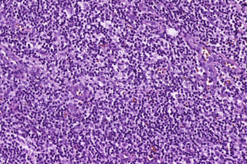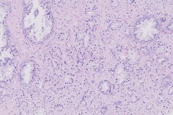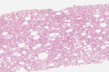
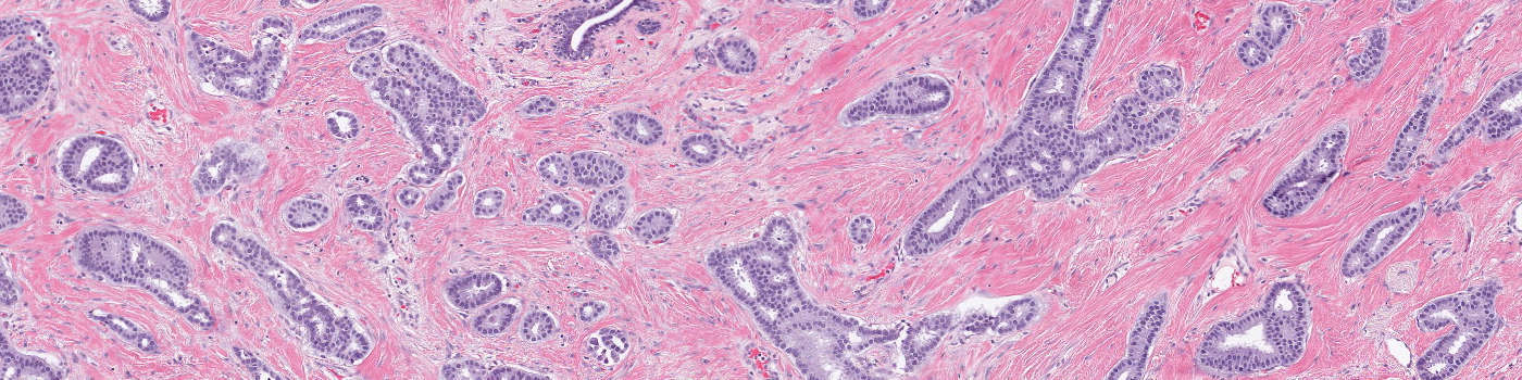
Raw whole slide H&E image.
#10093
Identifying tumor tissue and determining the percentage of tumor on Haematoxylin and Eosin (H&E) stained slides from formalin fixed, paraffin embedded (FFPE) tissue, has become one of the main methods for assessing sample quality.
With the “H&E, Tumor Load” APP an automated and standardized workflow for tumor identification on H&E slides can be introduced to the laboratory. The APP clearly outlines the tumor tissue and outputs the percentage of tumor present on the slide. This gives the user an easy overview of the identified tumor.
Raw whole slide H&E image.
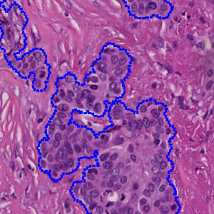
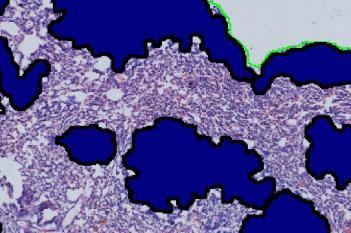
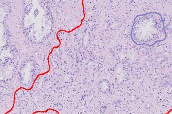
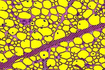
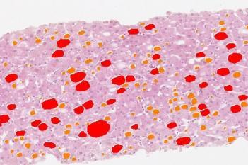
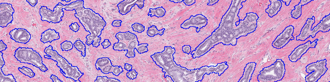
The output obtained from the “H&E, Tumor Load” APP. Tumor tissue is outlined with the blue ROI, which gives the user an easy overview of where there is tumor present on the slide.
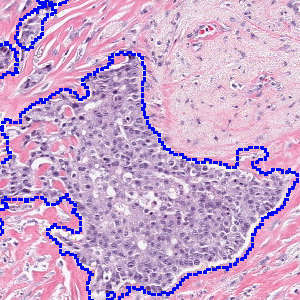
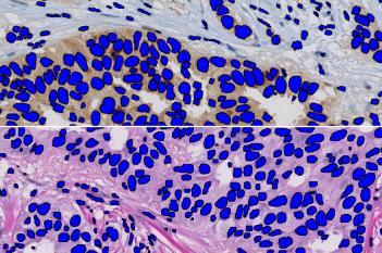
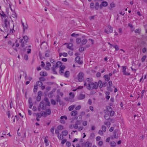
Zoom in on one of the identified tumor structures.
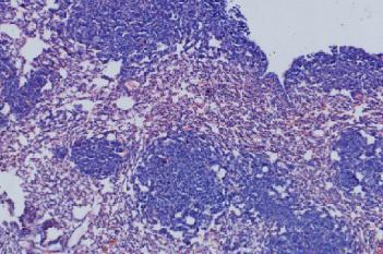
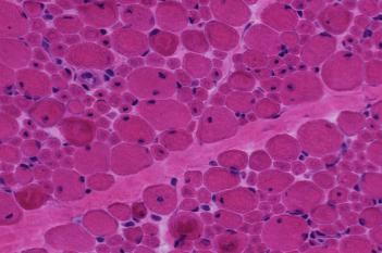
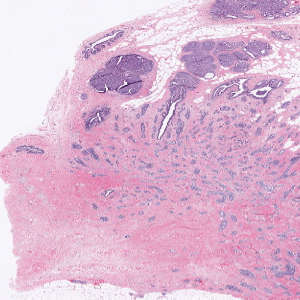
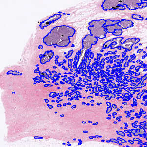
Raw image of a another H&E stained slide. The APP can be calibrated to different types of H&E staining.
Auxiliary APPs
APP: “01 Tissue Detect”
The APP “01 Tissue Detect” automatically detects and outlines the tissue on slide and limits the subsequent analysis to be within this region.
Quantitative Output variables
The output variables obtained from this protocol are:
Workflow
Step 1: Load and run the APP “01 Tissue Detect” for automatic outline of the tissue on slide.
Step 2: Load and run the APP “02 Tumor Load”. This APP outlines the tumor tissue with a blue ROI and outputs the tumor load.
Methods
The APP operates at a low magnification which enables analysis of a whole slide image within 5 minutes. For separation of the tumor and non-tumor tissue advanced input features, that are especially robust towards the staining variation inherent to H&E images, are used. The features are input to the decision forest classifier, which is useful for classification of images with varying staining intensity. The initial tumor and non-tumor separation is then refined by use of morphological and contextual post-processsing steps, and the identified tumor areas are marked by a region of interest (ROI). Finally, the tumor load, i.e. the percentage of tumor present on the slide, can be calculated.
Staining Protocol
There is no staining protocol available.
Keywords
H&E, Tumor Load, breast cancer, tumor detection, image analysis, decision forest
References
LITERATURE
There are currently no references.