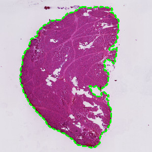
The relevant tissue of the whole slide image is automatically detected using “01 Tissue Detection”.
#10164
Developed for Medical College of Wisconsin
Centronuclear myopathy is a disease group characterized by the location of nuclei in the center of skeletal muscle fibers instead of the normal peripheral location. Symptoms of the disease include muscle weakness and diminished muscle tone.
This APP analyzes H&E stained skeletal muscle tissue to detect muscle fibers and nuclei located centrally within the fibers. The APP calculates the proportion of fibers with centronuclear myocytes.
The relevant tissue of the whole slide image is automatically detected using “01 Tissue Detection”.
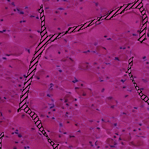
The muscle fibers are arranged in bundles separated by connective tissue. The protocol “02 Bundle Detection” automatically exclude the connective tissue from further analysis.
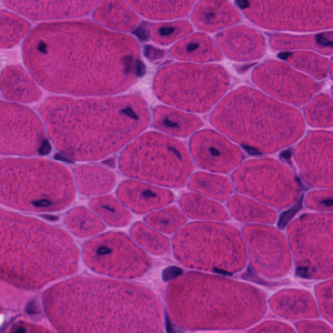
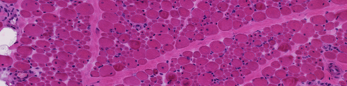
H&E stained skeletal muscle fibers. Some of the fibers have peripheral nuclei and some have central nuclei.
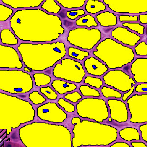
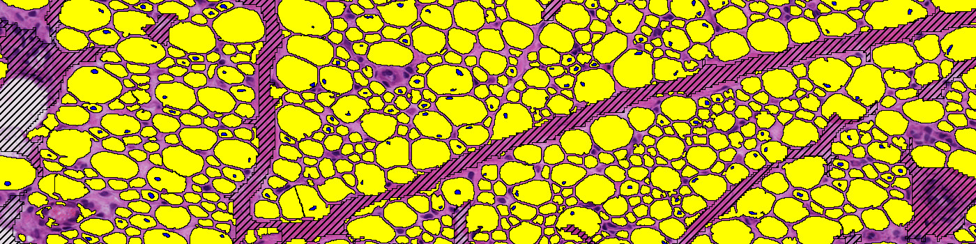
Detection of muscle fibers (yellow) and central nuclei (blue). Peripheral nuclei are included in the muscle fibers (yellow).
Quantitative Output variables
The output variables obtained from this protocol are:
Workflow
Step 1: Load and run the APP “01 Tissue Detection” for detection of the tissue in the slide
Step 2: Load and run the APP “02 Bundle Detection” for detection of bundles in the tissue
Step 3: Load and run the APP “03 Central Nuclei Fibers” for detection of muscle fibers and central nuclei
Methods
The APP consists of two protocols. The first protocol excludes non-myocytes (e.g. connective tissue) from the subsequent analysis. The second protocol detects and segments myocytes and nuclei. The myocytes are identified based on the H&E color band in VIS. A myocyte candidate is defined as an actual myocyte if it passes certain morphological measures based on circularity and area. Nuclei candidates are defined as actual centrally located nuclei if they are fully surrounded by detected myocytes.
Staining Protocol
There is no staining protocol available.
Additional information
An auxiliary APP “01 Tissue Detection” is included for automatic detection of the tissue in the slide.
Keywords
H&E, Centronuclear Myopathy, Skeletal Muscle, Image Analysis, Digital Pathology