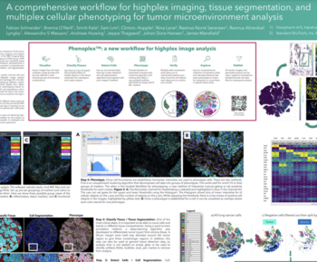
Imaging Mass Cytometry (IMC ) technology is a multiplexed imaging technique that generates high-dimensional spatial data at subcellular resolution without the complications of autofluorescence and cyclic imaging. IMC technology has two distinct whole slide imaging (WSI) modes: Preview Mode (PM) and Tissue Mode (TM). PM rapidly scans stained tissue to provide a comprehensive overview within minutes, while TM provides fast acquisition of the entire tissue at 5-micron resolution, mapping out the distribution of over 40 markers and revealing tissue heterogeneity. Both WSI modes enable researchers to make informed decisions about selecting tissue areas that warrant closer examination at single-cell resolution. Following PM, regions of interest (ROIs) are selected on the same slide for high-resolution imaging using Cell Mode (CM). This facilitates single-cell analysis of the ROIs identified during PM. These imaging modes together with an automated slide loader function support nonstop acquisition of tissue samples.
Smriti Kala1, Katherine Hales2, David Mason2, James Mansfield1, Regan Baird2, Hiroyuki Suzuki3
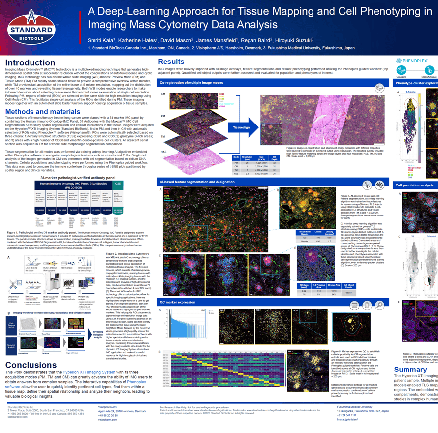
- Standard BioTools Canada Inc., Markham, ON, Canada
- Visiopharm A/S, Hørsholm, Denmark
- Fukushima Medical University, Fukushima, Japan