Get ready for an exciting journey into the world of tissue imaging with the first episode of our mini series. Dr Aleksandra Zuraw is joined by expert Regan Baird, our Scientific Consulting Sales Manager, to dive into the fascinating topic of multiplex staining. Find out the different modalities, what’s best suited for tissue image analysis and why, and the many benefits and uses of multiplex staining. Regan shares his expertise and insights to help you get the most out of your imaging modalities.
Further reading:
After his postdoc at Beth Israel Deaconess Medical Center in Boston, where he experimented with multidimensional, multimarker and multicolor single cell imaging modalities, 2D images of tissue stained with hematoxylin and eosin (H&E) seemed simplistic to Regan Baird, PhD. Tissue image analysis (IA) can be challenging when relying solely on H&E. So, multiplex staining was implemented to simplify the process and extract more information.
In this miniseries, Regan introduces us to the concepts of multiplexing and cell phenotyping and relevant image analysis approaches for multiplex data analysis. Multiplexing in life sciences refers to taking multiple measurements on the same specimen at the same time. The easiest method of multiplexing with tissue slides is IHC-based virtual multiplexing.
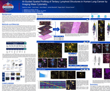
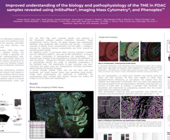
More precise methods of visualizing cellular colocalization of biomarkers include multicolor bright field IHC (up to five biomarkers per tissue, two biomarkers per cell), IF with spectral unmixing (up to nine biomarkers per tissue and cell), and imaging mass cytometry (up to 60 biomarkers in a single tissue section). The choice of method should be based on the experiment and scientific/diagnostic questions.
IF multiplexing is widely used for cell phenotyping in tissue, allowing for characterization of single cells and investigation of spatial relationships between cell populations. This provides information about the interactome of different cells and their environment.
- Let us know what you think of the episode and any topics you would like us to cover in the future by sending us an email at: podcasts@visiopharm.com