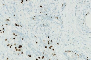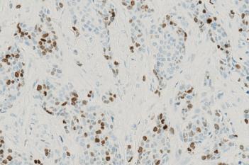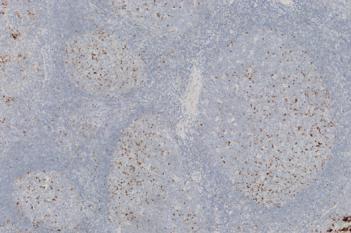
Breast tissue before this APP has been run.

#10187
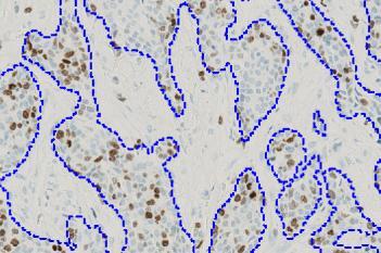
This Quickstart APP detects and outlines tissue in a brightfield (RGB) image without any manual annotation. Trained on IHC and H&E slides, it’s designed to get you started on tissue analysis without building from scratch. After tissue detection, you may segment specific regions of tissue you want to analyze further.
If you are working with tumor tissue the IHC Tumor Detection APP can quickly segment tumor regions. Once the region is selected, the Nuclei DAB Intensity Quantification of Nuclei DAB Quantification APPs can be run. Explore related APPs for downstream analysis below.
As with all of our Quickstart APPs, your image analysis can be scaled by queueing APPs to run in the background on an unlimited number of images—this is possible with the “batch analysis” feature available on both Discovery and Phenoplex™.
Working with multimodal data sets or TMAs? No problem. Run this APP after using the Tissuealign™ feature to align your images at the cellular level, or the Tissuearray™ feature to de-array your tissue cores.
Breast tissue before this APP has been run.
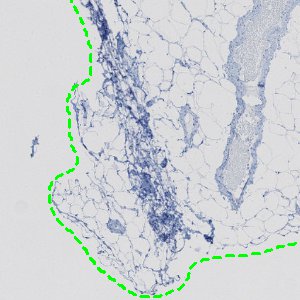
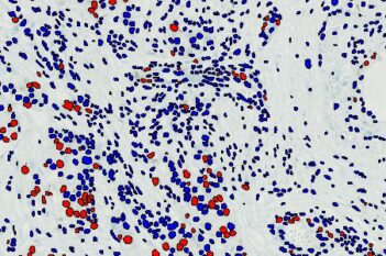
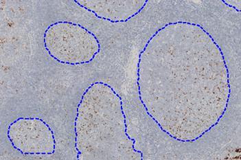
Outlined tissue with one click.
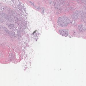
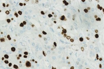
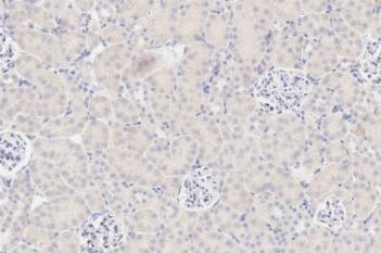
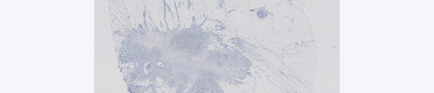
H&E image pre-processed.
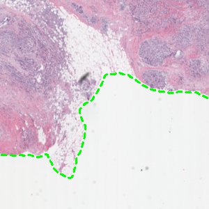
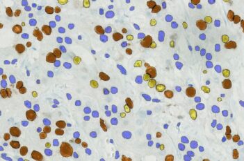
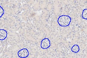
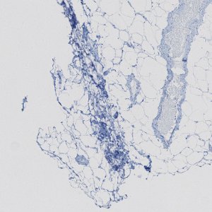
Tissue successfully separated from background in H&E image.
Quantitative Output variables
Output variables include:
Workflow