
Advancing Preclinical Lung Research with Automated Image Analysis: Interview with Simone de Meo, from Chiesi Farmaceutici
Simone De Meo
I am currently an Associate Scientist on a maternity cover assignment within the Neonatology and Pulmonary Rare Disease Preclinical Experimental Pharmacology & Translational Science Unit at Chiesi Farmaceutici S.p.A.. Recently, I completed my P.h.D. in Life Sciences at the University of Siena in collaboration with Chiesi Farmaceutici. Over the past years, I developed strong expertise in histological analysis, contributing to the characterization of animal models of chronic pulmonary diseases using both conventional techniques and Visiopharm software.
Can you tell us about your background and the work you do at Chiesi?
I have just finalized my PhD in the Neonatology Unit at Chiesi Farmaceutici. My research focuses on a preclinical animal model of bronchopulmonary dysplasia (BPD), which is the most common long‑term respiratory complication associated with preterm birth. Using this model, we aim to replicate the structural and functional abnormalities typically observed in premature infants who develop BPD.
A key objective of our work is to evaluate novel therapeutic strategies. At present, there are no specific pharmacological treatments available for this disease in preterm infants. Animal models therefore play a critical role, not only in improving our understanding of the disease mechanisms underlying BPD, but also in enabling the preclinical testing of potential new therapies.
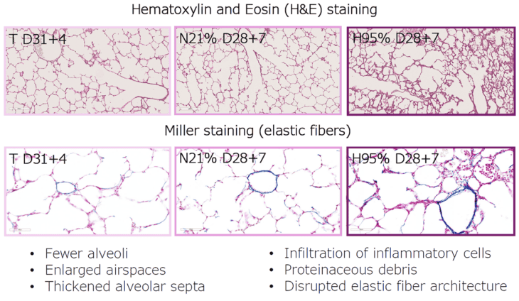
From poster: Functional and Histological Characterization of a Preterm Rabbit Model of Bronchopulmonary Dysplasia Exposed to Hyperoxia, Poster link
What type of analysis do you perform using Visiopharm?
During my academic training at the University of Siena, I developed strong expertise in histological techniques. When I joined Chiesi Farmaceutici approximately two years ago, I implemented these approaches here, focusing on immunofluorescence (IF) and immunohistochemistry (IHC) analyses of lung samples from our BPD animal model.
This particular model is technically challenging. During the first year of my PhD, I had focused on validating a wide range of antibodies for IHC and IF, which I then implemented using automated immunostaining protocols. In parallel, I began working with Visiopharm to develop image analysis applications capable of quantitatively assessing the signals observed in lung tissue sections.
Previously, I had extensive experience with manual morphometric analysis. While these methods are well established and widely validated in the scientific literature, they are often operator‑dependent and can suffer from limited reproducibility. By automating the analysis with Visiopharm, we have been able to significantly improve both the reproducibility and robustness of our histological data.
“Overall, automated morphometric analysis with Visiopharm has proven to be faster, more reproducible, and less operator‑dependent than manual approaches, substantially increasing the robustness of our data.”
How did you become familiar with Visiopharm, and how was the initial learning process?
Visiopharm was already in use in other research groups of my department within the company. At the beginning, I worked closely with a Visiopharm support specialist, who provided guidance on how to translate my initial ideas into feasible and effective analysis applications. This was particularly valuable, as not all conceptual approaches are directly applicable to our specific animal model.
Discussions with colleagues who were already using the software were also very helpful. In addition, we participated in a joint workshop with Visiopharm that brought together all users within Chiesi. This collaborative environment significantly accelerated the learning process. Over time, I developed multiple applications for IHC and hematoxylin & eosin (H&E)-stained lung sections, including approaches based on deep learning–based tissue classification.
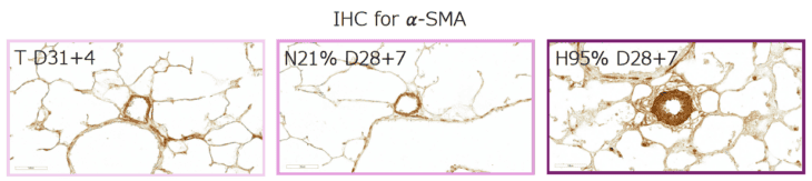
Postnatal exposure to hyperoxia results in a significant increase in thickness of media tunica of small pulmonary blood vessels (major axis length 30-100 μm); from: Poster link
What have been the main benefits of using Visiopharm in your work?
We initially worked with dark‑field imaging and later transitioned to bright‑field analysis for quantitative measurements. Currently, we are exploring a return to dark‑field approaches, including the use of RNAScope assays, to develop applications that quantify RNA molecules at the tissue level. This allows us to assess molecular differences between experimental conditions.
One challenge we routinely face is variability across experimental conditions, including differences in staining background between treated and untreated animals, or between distinct experimental setups. For each condition, applications require careful refinement of the training data to ensure accurate signal detection. Visiopharm makes this refinement process straightforward and flexible, which is essential for our work.
We have also developed applications for hematoxylin and eosin (H&E)–stained sections, using the same conceptual framework as our previous manual morphometric analyses, which served as our historical gold standard. By validating automated results against these manual methods, we established an integrative workflow that combines established techniques with automated image analysis.
Overall, automated morphometric analysis with Visiopharm has proven to be faster, more reproducible, and less operator‑dependent than manual approaches, substantially increasing the robustness of our data. We are now applying these automated workflows consistently across preclinical and clinical BPD studies, ensuring methodological consistency and enabling more reliable comparisons across experiments.
See the posterFunctional and Histological Characterization of a Preterm Rabbit Model of Bronchopulmonary Dysplasia Exposed to Hyperoxia
Simone De Meo, Chiara Catozzi, Matteo Storti, Giorgio Aquila, Enrica Scalera, Aurora Radicati, Carlotta Boggi, Francesca Ravanetti, Luisa Ragionieri, Roberta Ciccimarra, Xabier Murgia, Francesca Ricci, Gino Villetti, Barbara Bartalesi, Monica Lucattelli
Share this article