Transcript
Just give it a one minute for everybody to join, and then we’ll get started.
All right, I think we can let’s start.
Hello everyone, welcome to this Research Rebellion webinar.
With me today I have Diego Pedroza from Baylor College of Medicine. He’s gonna introduce himself in short. I just want to give you a little some instructions about today’s webinar.
So his presentation should take around thirty minutes and then there will be a Q and A at the end.
You can type your questions in the chat and we’ll take a look at them.
We will start now with just some questions to engage with you and to get to know you better. So let’s start with the first one.
Let me see if you can see it, actually.
What is your research area?
Yeah, sure. So, thank you guys so much for giving me the opportunity to talk. So, my research area primarily is directly with triple negative breast cancers, but it expands to lung and liver metastases, and it encompasses the usage of novel technologies like spatial proteomics. So because of that, basically I’m able to look at the tumor immune microenvironment.
So looking at breast cancer and studying the immune landscape of TNBCs basically.
Thank you, Diego. I can see that you are also answering right now. The majority is working on translational research and then preclinical drug discovery. I’m gonna move to the next question now. Let me see here.
Are you doing spatial analysis?
Yeah, of course. You know, as you’ll see in this presentation, we’re doing, we started doing spatial proteomics.
In this case, we did imaging mass cytometry.
We used, of course, Visiopharm’s, Phenoplex to analyze a lot of the data.
And now we’re actually moving towards spatial transcriptomics. I think a lot of the field is moving towards incorporating both transcriptomics and proteomics together to study the microenvironment in a whole tissue landscape.
Yeah.
Cool.
Let’s move to the next question now. So actually, majority of people are doing spatial analysis, so I think your talk is going be very relevant today, but also for those who are not doing, can get some inspiration.
Next one.
Do you have a multiplex instrument in house, in your lab?
Yeah, so actually, Baylor College of Medicine has a really good core.
I should say we have a lot of good cores.
So I don’t have one in my lab directly, but the cytometry core has two Hyperion IMC images, I should say, imaging mass cytometers.
And we actually just wrote another S10 grant to get a new COMET spatial proteomics instrument. And then the single cell genomics core has a MuirFISH platform as well as a 10x Genomics spatial transcriptomics platform with Xenium and Visium capabilities.
Wow. You guys are well equipped.
Well, you know, I was just in San Diego, and 10x Genomics is coming out with the Terra now. So I feel like we’re always at the end of a we’re always lagging behind. So, these facial platforms are always coming up with new things.
That’s true. But it’s also exciting, right?
Cool.
Most of people today here don’t have it in house. So again, another opportunity to learn the capabilities of I and C today. I think that’s going to be your focus, right?
Yep.
And then last question.
Are you currently using Visiopharm software?
Yeah. Yeah. Like I said, you know, we we did use a lot of we actually use Visiopharm a lot. That was the in house software that we used.
And we actually I was actually trained, and I’ll talk about it here. We use some type of machine learning algorithm.
But now we actually have the new AI platform, which I have not used yet, but it is available here at Baylor, and people are being trained on it. So I I would have I have not used a new AI platform, but I plan to be trained on it soon.
So maybe you guys have a better understanding of that one than I do.
Let’s see. Alright. So I think we got some insights here, and I will hand it over to you, Diego.
Let me just put your presentation here in on the stage. I think everybody can see now, so feel free to start it.
Great.
Well, thank you everybody. Again, my name is Diego Pedroza. I’m an instructor at Baylor College of Medicine. I’m in the Department of Molecular and Cellular Biology, and the talk of today will be cellular networks that lead to intratumoral heterogeneity associated resistance in metastatic triple negative breast cancers.
So I have no financial relationships to disclose, and the monoclonal antibody that we use was actually kindly provided by Syndax.
So I just want to start off with some brief breast cancer statistics. I know not everybody works on cancer or breast cancer for that matter.
Breast cancer has remained the highest and most commonly diagnosed type of cancer in women.
And as you can see, up to forty two thousand women are estimated to die of breast cancer in twenty twenty six.
And over the past several decades, again, breast cancer has remained the most diagnosed type of cancer in women, just over lung cancer as well.
There are several types of subtypes of breast cancer, some that are more aggressive than others, some of them being luminal A, normal like luminal B. So these are what are called hormone receptor positive. So this graphic is kind of deceiving. You see best prognosis, but we don’t really want to say that.
Any prognosis is not a good prognosis, But these are the more treatable ones, I would say. So the hormone receptor positive are more treatable. They have estrogen receptor or progesterone receptor positive. So we have a lot of drugs that can actually target ER, PR.
Then you have HER2 enriched. Now there are a lot of other drugs that can actually target HER2, a lot of ADCs now, like antibody drug conjugates that can actually target HER2, which are now FDA approved.
And then we have triple negative, which is actually the worst of all the breast cancers, basically because it lacks all the receptors, right? So it lacks ER, PR, and HER2, so we don’t have any targets.
And therefore we can’t give any of the treatments for any other of the breast cancers.
So, you know, our study basically spans a lot of clinical trials, and so one of the most famous trials was here done across the street from us at MD Anderson. This was called the ARTEMIS clinical trial, And so basically what they wanted to see was they wanted to see what happened to patients after these patients received what’s called neoadjuvant chemotherapy.
So patients presented to the clinic with breast tumors, they were given chemotherapy, and they had tumor profilings before and after chemotherapy. And essentially what they ended up seeing was in the patients that the chemotherapy became resistant or where the chemotherapy failed the patient, they had these upregulated pathways that were associated with epithelial to mesenchymal transition or EMT, as well as some immune signatures like monocytes and macrophages.
And so they basically had the patients that didn’t do well following chemotherapy had this high EMT, high immune signature, which is marked by high macrophage scores.
And then we went across looking for other things in the literature because we were very intrigued by these macrophages that were being observed about five years ago.
And we came across this manuscript that was published in Nature Communications several years back now, where they actually looked at several in silico based analysis, some that we all use like MetaBrick, TCGA, and basically you can see that they were able to cluster these. So clustering and cluster C basically span patients that did well following certain treatments, and you can basically see that in cluster A, for example, or sorry, cluster C, for example, you see a lot of immune signatures, very good immune signatures like B cells, T cells, plasma cells, etcetera. Contrary to that, they actually saw patients that landed in cluster B, and these clusters actually had very high levels of macrophages.
And in this case, they were these M2 like macrophages. Or at the time, the nomenclature was just to call them M1 and M2. And so these were called M2 like macrophages. And again, you can see cluster B basically, you know, they cluster with these gamma delta T cells, which don’t favor or have worse prognosis relative to patients that have high T cell abundance that do better in treatment response.
So to take advantage of this, our lab actually had engineered genetically or actually had engineered or genetically engineered mouse models.
These are Balb C mice. These have an immune system. And so we were basically able to take the mammary gland and transplant these and we were able to generate a transplantable bank. These mouse models spanned it from basal like, luminal and cloud and low. And because they have an immune system, can look at the different immune cells. So for example here we can look at F-four eighty cells so you can see the different types of macrophage levels that these mice have, as well as neutrophils. And so you can see, for example, the D122151R, these mice have very high levels of macrophages contrary to where the other ones have high levels of neutrophils.
We were able to expand this to more models as well and then do some basic transcriptomic profiling. And we were able to see that these macrophage enriched subtypes, these mice that have elevated levels of macrophages at this very high level of EMT just like what was seen in the ARTEMIS trial in the patients.
So this was very exciting to us because now we actually had relevant mouse models that basically shared what was being observed in the patient population.
So this is I think where it gets pretty exciting. So this is where we were able to use the workflow for Imaging Mass CyTOF.
So I will give credit where credit is due. So we partnered with Doctor. Weigu Wu and Pop Order. So they are the ones that run the core and they were very instrumental in helping us develop the panel and testing all these tissues and setting up IMC because when we got here it was still fairly new.
And so you can see we get some very beautiful images here. These are of our mouse models.
And so you can see the green here denotes the amount of macrophages, with white being Ki67.
So the majority of it is macrophages and proliferating tumor cells.
And basically, aside from the levels of immune cells, what changes is the architecture of tissue.
So you can see that the basal like here, these two, actually have a very good basal compartment compared to the lack of architecture here. So the ones that have this very high macrophage EMT don’t have that nice tight junction grouping, so they’re very disorganized tissue. So they’re extremely, extremely aggressive tumors. And then again, we were able to, of course, quantitate this, so you can see these have various degrees of macrophages throughout the tissue.
Of course, we’re an imaging lab, so we always confirm this. So we went ahead and did double immunofluorescence, and so with the red being macrophages and the green neutrophils.
And again, you can see a lot of red here in a lot of these models with some of them having various degrees of neutrophils and macrophages and some just being rich in macrophages.
And again, you can see the very nice architecture here where in this model you can see just the macrophages running through certain compartments of the tissue.
Not so much, just disorganized like it is in these models here. And again, in the 228L model here, the more basal like you can see more neutrophils compared to the cloud and low models.
So it became apparent to us that these macrophages were playing a role in the development or at least resistance in cancers. So these tumor associated macrophages, again, they have different levels of markers, so they can be denoted by CD11B, CSF1R, or F480, which I just showed you. But they play a lot of different roles. They can play a role in immune regulation, inflammation, angiogenesis, as well as stem cell maintenance, therapy resistance, intravazation, and as well as metastases. And these were some of the early markers that were captured using flow cytometry, and now with spatial single cell sequencing, we’ve expanded these markers to basically hundreds of markers now to denote various levels of macrophages.
And over the years, it’s not just been us. There have been other labs that have also been interested in targeting macrophages. So this is a lab, Jennifer Guerrero from Dana Farber, where they try to target macrophages in combination with PARP inhibitors.
Mikaela Eglobat’s lab, who’s now at Johns Hopkins, who was able to reprogram them rather than completely destroying them like we do.
Even in Lev Becker’s lab where they actually use nano devices to target them at University of Chicago.
And then our previous publication where we use a small molecule inhibitor to completely destroy them in combination with chemotherapy and breast cancers.
Unfortunately for us, the small molecule inhibitor went to clinical trial and it failed in the I SPY two because of liver toxicity, so this drug was no longer used for the clinic to treat breast cancers.
So fortunately though, we were able to outsource a different way to target macrophages.
In this case we use a drug or an antibody called axatilamab. So this is a monoclonal antibody that can bind to a surface receptor, in this case CSF1R, and it can directly bind to CSF1R impeding the activation or downstream signaling of the CSF-1R receptor.
And this is called Nictimbo. I don’t know in the UK or in Europe, but here I’m sure they’ve shown commercials for it and we have not profited from anything out of this. I don’t know who is gonna profit from this, but not us. But Nectivo or axatilamab is currently approved for chronic graft versus host disease.
So we were able to actually use it and repurpose it pretty quickly, which is pretty great for us.
So we took this initially, we put this into a mouse model. We use it in a bulbs seed background. We can transplant the mouse models. We wait about a week and a half. These mice have palpable tumors by then. And then we randomize the mice.
We give the treatment four weekly treatments with, immunostimulatory cyclophosphamide, again just once a week. And then we stop the treatment after four weekly treatments at day twenty eight and then we assess recurrence.
And we did this across different mouse models with the various different levels of macrophages. And so you can see that several of these mice have very, very good response even after we stop treatment.
Some don’t have that good response, you know, they have partial response. And some that are actually more enriched than neutrophils don’t have any good response.
So we took the full responders and we wanted to see what was really the mechanism behind this. So we proceeded to initially do single cell RNA sequencing. We actually have a method where we can pull them together and do cell hashing to remove the batch effect and cost of this.
And again, we can do a single cell and you can see our IgG control. We have a large population of macrophage monocytes. The monoclonal antibody depletes this population pretty efficiently. Chemotherapy itself will increase to a certain degree does have a level of inflammation. So we do see a population of macrophages, but the combination does improve this and we see a huge increase in T cell abundance, as well as B cell abundance, which led to the elimination of a lot of tumor cells. We also see again, as I mentioned before, a large accumulation of different types of macrophages that we transcriptomically have profiled now.
We can also do signaling pathway network analysis where the combination basically impedes now direct communication between the tumor cell and the macrophages and monocytes.
We also go ahead and do what’s called tumor cell rechallenge. So in the mice that completely eliminate the tumors, we actually take fresh tumor cells and re implant tumor cells into the mice to rechallenge their innate and adaptive immune system.
And again, you can see that some of the mice completely reject the tumor cells that we implant.
And because they don’t have any more tumor cells, we are able to actually remove the mammary glands from them.
We can do H and E staining and we can look for immune cells so we can detect B cells as well as B cells with T cells coming into close contact.
We can do plasma analysis to see what’s going on and we of course detect high levels of interferon gamma, IL-five and M CSF, which is our biomarker because M CSF is no longer being able to be internalized. Therefore, we can say with confidence that the drug is working pretty well. This is a cytokine representation of what we believe is going on and just for the interest of time, I won’t get into it, but this is all published, so I’m happy for anybody to go read. So this is just the first conclusion to our first aim here, which was using primary tumors. We did see increased CXCL9, which is now what the trend is in macrophage biology. We see increased interferon gamma and M CSF, which we think would be prognostic markers.
So we do think that the combination of our monoclonal antibody with chemotherapy did lead to a good prognosis in primary tumors.
But we’re interested in actually treating metastatic breast cancers.
So as you know, when breast cancers or any type of cancer metastasizes, that’s when the five year relative survival dramatically decreases. And unfortunately for breast cancer, the reality is that the disease remains fatal for up to ninety seven percent to ninety nine percent of those diagnosed.
So to combat this, what we do is we actually do tail vein injections and we actually can generate established metastases. This is generated by HNE staining. We inject BRDU. BRDU is an intercalating agent, so it’ll basically bind to DNA And so you can detect it by immunofluorescence, so we know that it’s actively binding to actively dividing cells. So we have established dividing cells in the lung before we start our treatment.
We repeated the same thing. We used imaging mass cytosol because we really wanted to see what was going on. And we identified these small little micro mets, micro mets one and micro mets two, because we saw a really good response.
And this led to some what we call intratumoral heterogeneity. So these two micro metastases, even though they were close to one another, they displayed various levels of proteins. So one of them had higher levels of PD-L1 and the other one had actually higher levels of imentin. And unfortunately this eventually led to one hundred percent recurrence because we stopped the treatment.
And as you can see here, the recurrence was due to the macrophages just came back with a theory and we had no T cells anymore.
So before I get into how we went about treating this, again, I just want to go through the workflow of what we were able to do with the IMC. So this is one of the ways that we were able to do it. So we were able to take one of these lungs that were treated, This is just using multiple channels. We can split into DAPI channel, which is a nuclear stain.
We can do cell segmentation using Visiopharm software. This is all with the Visiopharm software, by the way.
We can split into individual channels like the Menten and four eighty. This is our EMT marker, macrophage marker proliferation marker.
And then we can also put them together with cell masking, which I think is really cool.
So if you were to zoom in here, you could actually see this is sort of populating with almost like a fake how do say, like a fake colors, if you will, based on the intensity levels here. But it’s really cool because it allows you to really hone in on the markers themselves.
And then we are, of course, able to do nuclear segmentation, marker masking, like I mentioned. We were able to obtain marker density, dimensional reduction. We were able to do neighborhood analysis and then overall we were able to measure the time, what we call the tumor immune microenvironment.
And so of course, this is just showing you in the lung, our antibody depletes all the macrophages. This is FR-eighty staining. This is a control lung where you can see we are able to split channels here. This is all the channels that we get. These are control lungs with the tumors. So vimentin, CD44, these are the macrophages with Cas67.
And then this is when we treat them with a combination. I’m not sure why it looks a little blurry, I’m sorry about that. But basically you can see we eliminate F480 here and Ki67 is able to go low.
But we do have some PD-L1.
And we were able to use Visiopharm to actually look and find elevated levels of PD-one and PD-L1 and using dimensional reduction analysis. So we were able to find that the mechanism of resistance here that these lung mets were exhibiting was increased de novo synthesis of PD-L1. So of course to combat that we bring in anti PD-1.
So we incorporate anti PD-1 now. You can see now compared to the treatments, we start to see more of a good lung.
And when we incorporate anti PD-1, we see less tumor burden. We apply imaging mass cytosol again. We get these beautiful images.
IMC is one of the most beautiful techniques I’ve ever used to get images. We can split the channels at just incredible resolution. And then using Visiopharm, we did neighborhood analysis, which allowed us to do distance analysis. And basically we were just able to show that in the tumors where we get a good response, where we have antitumor immunity, we see CD4, CD8 and neutrophils coming in where we see this tumor cell killing contrary to where we have these high macrophage abundance with CD4, CD8 and neutrophils being distant from the tumor cells themselves.
Yeah, and so this was the long term response in the lung. As you can see again, once we repeated the experiment, we were now able to prolong the response of these mice up to seventy days.
We didn’t see any drop in weight and we were able to rechallenge these mice and we saw a really good response in the mouse model.
We look for tertiary lymphoid structures which in the literature have shown to be a good source of response in human samples. And again, using double immunofluorescence, we’re able to now capture, you know, these CD20, CD8, B cell, T cell lymphoid aggregates coming together, which we think we’re able to eliminate the tumor cells.
Just really quickly because I know I’m running out of time, we were able to obtain with our collaborator Chuck Perru from North Carolina data from the AURORA clinical trial where they were able to basically show that in patients that had metastatic disease, you know, liver actually had the lowest immune signatures. So triple negative breast cancers that went to the liver for some reason had reduced immune checkpoint blockade.
And this was also showed by Bowden Miller’s lab, which I think you guys should be familiar with Bowden Miller because he’s kind of like the grandfather of using the spatial proteomic techniques and we’ve actually used a lot of his pipelines to analyze a lot of the data.
And so he actually, his lab actually also saw that patients that had liver metastases had exhausted CD8 T cells and they actually had these arginase one positive myeloid populations, which we now know are immune immunosuppressive. So we decided to test this in our models, so we generated liver metastases. So you can see here these liver mets are immunosuppressive, so they have no T cells in them but they’re loaded with macrophages and neutrophils. We proceeded to test this as well. We have established metastases before we start our treatment. This is just showing after we do our treatment, you can see the liver architecture improves significantly. And again, we can drop the levels of tumor cells in the liver when we start our treatment now including immune checkpoint blockade.
We applied imaging mass cytometry and again, you can just see some beautiful images here where when we give our triple combination, we now start to see this immune microenvironment come to life with decreased macrophages and decreased Ki67.
And then most importantly, you know, for us was the fact that the antibody doesn’t actually deplete the normal macrophages. It only depletes the tumor associated macrophages that are directly within the tumor bed and not in the residual stroma of the liver, which is important.
We repeated this and now we were able to show that we could prolong the survival of these mice up to fifty six days. We also observed no weight loss.
We re challenged the mice and here we see one hundred percent response.
So all the mice rejected the tumor cell re challenge.
We did an adopted T cell transfer. So we took a spleen from these mice, we removed the T cells, and we directly put them into mice that had actively growing tumors in them with anti PD-1. And you can see that these mice slowed the growth of these tumors and the mice that actually received these T cells actually had decreased spleens, which is actually an indicator for good response.
And these mice also had increased CD8 T cells in both the spleen and adopted T cell tumors themselves.
Again, looking at Aurora samples themselves, what we were able to find was that the patients that had these tumors in the liver and the lung actually had increased CSF-1R as well as MRC1 and TREM2 positive macrophages.
And so just want to conclude here because I know I’m a little bit over time, is that in our primary tumors, our combination treatment where we see a complete response, we see increase in certain cytokines including interferon gamma and CXCL9 with decreased tumor associated macrophage signatures such as CSF-one hundred and TREM2. In the lung metastases, we initially see intratumoral heterogeneity that is associated with resistance.
And so to overcome this resistance we had to add anti PD-1 where we now observe these tertiary lymphoid structures. And in the liver we of course observed increased adaptive immunity by seeing increased B cells with CD8 T cells as well as CD4 neutrophils coming together.
But of course the most exciting part for us is that this has now led to the start of a clinical trial. So this clinical trial has now been ongoing for about six months. So this is called the New Start Phase Ib clinical trial. So this is a cyclophosphamide, axatilamab with retifanlimab.
This is an anti PD-1, generic anti PD-1 drug. So this is essentially now being given to patients with triple negative breast cancers that are metastatic or that have metastasized to the lung and the liver. And this is being done at MD Anderson by Doctor. Boralim.
And so we’re actually now in the process of harvesting or collecting both plasma samples to do the cytokine as well as biopsies to do the spatial analysis, which we will do IMC as well as spatial transcriptomics now.
And so with that, I just want to thank everybody in the Rosen lab, Sean Zhang’s lab, as well as everybody in the Cytometry and Cell Sorting Core, especially Weigu and Paul for helping me, all of our collaborators, as well as our funding.
And then I just want to say hopefully, I will be starting my own lab in October, so if there are any students out there or post docs, hopefully if you guys want to come to Houston or wherever I go, be sure to look out for applications.
Wow, very exciting. Congratulations on the new love coming. We can actually we are recording this call, and we will send the recording to all the that signed up. So we can actually mention that on the email that you are open for applications.
I’m sure we’ll have people interested.
Well, thanks a lot for sharing all this work that you have done. Very impressive. I’m sure you could spend the whole day talking about it.
You mentioned, mean, it’s also very exciting to see years of research becoming a potential treatment, right?
Going to clinical trials, very exciting.
The spatial analysis that you mentioned, is it also going to be part of the clinical trial?
Yeah. So we actually put in a SPORE grant.
We’re hoping it’ll be funded in October.
And we are looking into either using the Imaging Mass Cytop or COMET platform.
And so we are in talks with Wego to use Visiopharm’s AI platform to use a cell segmentation for that, because we’re going to do the, you know, we’re going to get biopsies from all these patients, and we want to see, you know, the, we want to make sure we can compare the patients that, you know, respond to the ones that don’t respond.
Yeah. That makes sense. And is there a reason why you decided to use imaging mass cytometry in your study?
Well, yeah.
So the reason is because we actually got a pilot grant to do it, and I was one of the first ones to test it and set it up.
They bought the instrument, and they were like, oh, we’re gonna give pilot grants.
We need help, basically. Because it took a lot, it almost takes a year long process to get the tissue. You know, we had to use spleen, normal mammary gland, tumor tissue, brain tissue, other types of tissues to find normal levels of these immune cells.
And then really look at different antibodies because you have to titrate the antibodies as well.
And then test all the antibodies, see which antibodies work. So yeah, it took a long time, but basically because the answer to your question is because the instrument was here, and they gave us the money to test it.
And so Yeah, okay. That makes some sense.
But now I think imaging mass cytosol once we tested it, the resolution is incorrect. I mean, yeah.
Cool. And, well, I can see I don’t know if you can see the chat, but I will read the question for you from Mass here.
So you mentioned that axitumi, you know, the name of the drug was used to treat GVHD where you want immunosuppression. How is the same antibody able to treat cancer where you want immune activation?
Yeah, I think that’s a great question. So basically, you know, so here we’re directly using it to target these macrophages, and so initially we weren’t sure if it was just gonna just target all the macrophages, monocytes, myeloid populations indiscriminately, and so, like some of the other drugs, like pexidarnatib did, but actually after a lot of testing, we found out that axatilamab actually will selectively deplete the tumor associated macrophages that highly express CSF-1R.
And now we have new evidence showing that it also depletes the ones that are TREM2 positive, which are immunosuppressive.
And the answer to the question is by itself, it actually won’t work very well. We have to give the combination of chemotherapy.
So the chemotherapy is the one that induces the, inflammation.
And then when we deplete the macrophages, that combination is sort of a magic that works really well, where we see the inflammation, T cells, B cells coming in together. And then when we deplete the macrophages, now the macrophages can’t get rid of the T cells anymore.
And so now we see an even bigger immune response that’s capable of eliminating the tumors.
Thank you. And Sahar wrote, Do patients exhibit elevated macrophage levels?
Yeah, so in the patients, you know, in the ARTEMIS clinical trial, those patients did see, the patients that became resistant to the clinical, the patients that became resistant to neoadjuvant chemotherapy transcriptomically had higher levels of macrophages. And then Clinton Yam at MD Anderson also showed that at the protein level, they also did immunofluorescence staining to show that the same patients also had higher levels of macrophages.
And now there’s a lot of evidence showing that patients that have metastatic samples, either in the liver or in the lung, have accumulation of macrophages.
And so that’s why we think that our therapy is beneficial to those patients.
Thank you. Tina, are there other drugs that target macrophages?
Yeah.
So the other drugs, you know, as I mentioned, pexidarnatib is the other drug that will completely destroy the macrophages that we just used.
Michaela Alcobat actually has really good ways of targeting macrophages. Not necessarily inhibiting, but I think reprogramming them is a different way of targeting them. So there are a lot of other drugs to target the macrophages.
So another question from Mes. When you analyze tumor microenvironments, are you looking at marginal zones or other subcompartments to compare immune cell population?
Yeah, when we actually do imaging mass cytoph, we actually do different regions of interest, so we can get different ROIs. So we just don’t look at the one different region, or one sorry. We don’t just look at one specific region, we actually get random regions.
And so therefore we kind of stitch the image together.
And we come up with, you know, a large compartment of regions to study the microenvironment that way. So we’re not just biasedly choosing one or two regions to analyze it.
Thank you.
Another question from Are the tertiary lymphoid structures, TLS, you observed associated with HEVS?
Also, do you see a difference in TLS before and after different treatments?
Yeah, so I’ll answer your second question. So, yeah, so the tertiary lymphoid structures, basically, we only see them after we give immune checkpoint blockade. And we only see them where the mice generated a complete response in the lung.
And interestingly, that’s one of the differences between the lung and the liver, is in the liver we don’t actually see these structures, lymphoid structures.
We see a different type of transcriptomic reprogramming of recovery of Cooper cells and progenitor cells.
But it seems that this tertiary lymphoid structure is a good prognostic marker in both humans and mice in lungs after immune checkpoint blockade. I’m not sure what you mean by H E B, sorry. If you can spell it out for me.
Yeah.
Oh, high intensity Yeah, yeah.
No, that’s great. Yeah, so actually we do. So if you do like CD31 staining, and you see, you know, where you see angiogenic or high blood vessels, we actually see a lot of immune cell compartments. So you do see a lot of blood vessel formation with a lot of tertiary structure formation there as well.
Cool.
I have a random question to you.
What are you the most excited about in the spatial biology field right now? Like, what do you see as, yeah, exciting things happening?
Yeah. Well, I’m equally excited and scared for AI.
You know, I have been I have been using several AI platforms that are capable of giving us a lot of information.
But the more I use them, the more it worries me because not necessarily, I think, in what people are worried about, that they’re gonna take our jobs.
But I think, unfortunately, we’re gonna see a lot of misinformation, I think, that’s gonna be published. I think a lot of the AI platforms don’t really spit out a lot of real information, and if people are using them, I would caution, please double check what you’re using.
So I was just talking to one of my students. We were actually using an AI platform, and, you know, it was feeding us a lot of information, and I was like, wow, this looks so great.
And then, you know, we ended up asking it to give out the the pipelines, and when we ran it ourselves, and we started encountering errors.
And so we were asking it again, like, why are we getting errors? And it was like, oh, yeah. You guys were right. You know, we I don’t know.
I don’t know why I ran it this way. And I was like, oh. Yeah. It’s you know?
But if you don’t actually, like, double check or something, it will just give you some of the stuff that you want to see. So I just caution against that.
Just be wary of that for everybody using it.
I’m excited for it because it does it can give you a lot of, lot of good information if used properly.
But also for spatially, like you mentioned, I think for us, the ability to study the microenvironment in both mice and humans is gonna be extremely important, and to overlap that. Because then for the clinical trial that we’re doing to capture the transcripts in those patients, for example, if we find transcripts that are not responders or proteins, then we can directly put them back into the mice and try to target them in the mice and see if we can overcome that resistance almost immediately for, and try to find predictors as well.
Cool.
So this is gonna be the continuation of your study now?
Yeah, and so that’s basically the basis for my lab moving forward, is incorporating a lot of spatial proteomics and transcriptomics for both human samples and mouse samples.
Oh, exciting. So if anyone here is interested in this topic, you should talk to Diego and see the opportunities there.
Yeah. Please read our paper and reach out anytime.
Yes. All right, thank you so much Diego. It was really nice to see your hard work, impressive work here today. And thanks everybody for joining.
We’ll share the recording this week and you can also wait for more information about our next webinar in August.
Awesome.
I’ll give everyone back your time and have a good end of the day or beginning of your day, depends where you are in the world. All right, bye bye. Thank you.
Thank you for having me.
How spatial biology in triple-negative breast cancer reveals the tumor microenvironment
Spatial biology in triple-negative breast cancer is helping researchers uncover the complex interactions between tumor cells and the immune system. In this webinar, Diego Pedroza from Baylor College of Medicine shares how spatial proteomics, imaging mass cytometry, and immunotherapy are advancing our understanding of treatment resistance, metastatic disease, and new therapeutic opportunities in triple-negative breast cancer (TNBC).
Drawing on advanced spatial biology techniques, including imaging mass cytometry, single-cell RNA sequencing, and multiplex tissue analysis, Diego demonstrates how tumor-associated macrophages contribute to therapy resistance, metastasis, and intratumoral heterogeneity in both primary and metastatic breast cancer. The webinar highlights the use of anti-CSF-1R therapy (axatilimab) combined with immunochemotherapy and immune checkpoint blockade, showing how this approach can activate adaptive immune responses, reduce tumor burden, and generate long-term anti-tumor immunity in preclinical models.
You will also learn how spatial analysis platforms such as Visiopharm enable researchers to map immune cell interactions, identify resistance mechanisms such as PD-L1 upregulation, and uncover clinically relevant biomarkers. The session further explores the translation of these findings into a newly launched Phase Ib clinical trial for metastatic triple-negative breast cancer, bridging preclinical discovery and patient treatment.
Key topics covered:
- Triple-negative breast cancer (TNBC) and treatment resistance
- Tumor-associated macrophages and the immune microenvironment
- Spatial proteomics and imaging mass cytometry (IMC)
- Single-cell and spatial transcriptomics workflows
- Immune checkpoint blockade and anti-CSF-1R therapy
- Biomarker discovery using multiplex tissue imaging
- Intratumoral heterogeneity in metastatic cancer
- Translational cancer research and clinical trial development
- Applications of AI and machine learning in spatial biology
This webinar is ideal for researchers working in oncology, immunology, spatial biology, translational medicine, drug discovery, and biomarker development, as well as anyone interested in the latest advances in spatial omics and cancer immunotherapy.
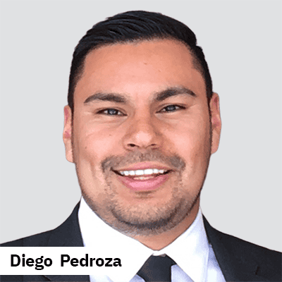
Diego Pedroza, PhD
Bachelor’s degree in Molecular and Cellular Biochemistry from the University of Texas El Paso, master’s in Biomedical Sciences from Texas Tech University Health Sciences Center (TTUHSC) El Paso and Ph.D. in Biochemistry, Cell and Molecular Biology from TTUHSC. Shortly after joining Baylor College of Medicine (BCM) I received aT32 postdoctoral fellowship from 2020-2022, applied and became an American Cancer Society Postdoctoral fellow from 2023-2024 and then transitioned to a K99/R00 before being promoted to Instructor in 2024 at BCM.