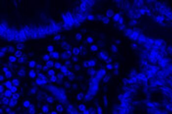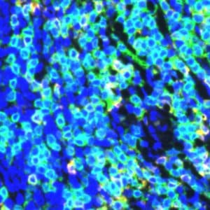
Raw fluorescence image with DAPI staining.
#10190
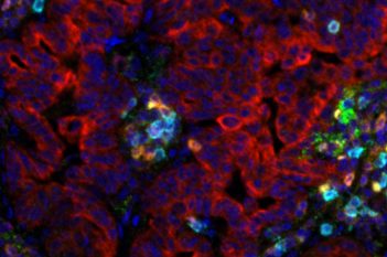
Easily segment your fluorescence images with a click. This Quickstart APP detects the nucleus with the DAPI channel and outlines the cell body, speeding up your analysis. Output variables include the total cell count and multiplexed intensity measurements for the nucleus, cell body, and entire cell.
As with all of our Quickstart APPs, your image analysis can be scaled by queueing APPs to run in the background on an unlimited number of images—this is possible with the “batch analysis” feature available on both Discovery and Phenoplex™.
Working with multimodal data sets or TMAs? No problem. Run this APP after using the Tissuealign™ feature to align your images at the cellular level, or the Tissuearray™ feature to de-array your tissue cores.
After analysis is complete, you may explore your results with the Phenoplex™ Guided Workflow or the Data Exploration and QC tool (available on Discovery and Phenoplex™).
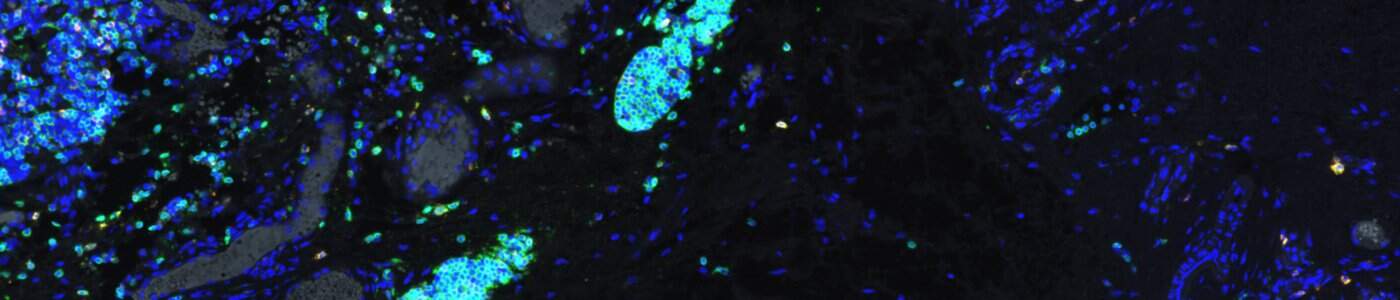
Raw fluorescence image with DAPI staining.
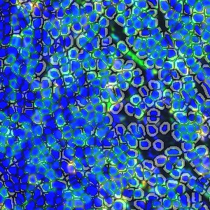
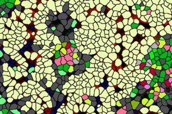
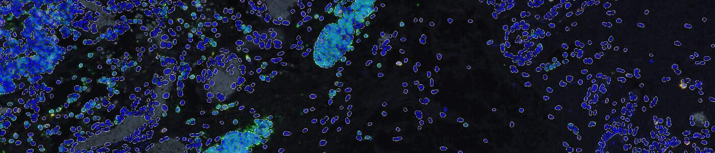
Cell segmentation.
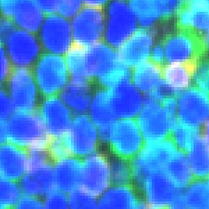
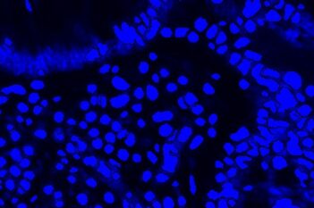
Zooming in on DAPI stained tissue.
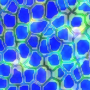
Zoomed in view of cell segmentation on DAPI stained tissue.
Quantitative Output variables
Workflow
After loading your image, follow these steps.
Step 1: Open the 10190 – Cell Detection, AI (Fluoresence) APP.
Step 2: Select a region of interest, or skip to next step.
Step 3: Click “Run APP.” A dialogue box will appear where you must set the input channel to match your DAPI channel. Once saved, the APP can be run with the batch analysis feature so you can keep working on other images.