Transcript
First of all, I want to thank you for giving me the possibility to present my data here. I’m Jose leader at this team.
And, and today, I’m gonna show you two different stories obviously related on digital pathology, and Visiopharm in this specific case. And, and I’m gonna show you, all the field of of blood vessel, tumor blood vessel that is actually my field. I’m an expert on on vascular biopsies.
So as I told you, the main focus of my research is to investigate how tumor blood vessel, are involved in in the tumor progression and automatically in the treatment of, of the tumor, different type of tumors.
And, I studied two different tumors. One is a breast cancer, the static breast cancer. And today, I’m gonna show you, this story on, on how the placebo normalize, blood vessels, that’s improving outcome outcome.
And then a second project in which, I show, and I demonstrate how glioma glioma cells caught, for existing blood vessels.
And, and now I’m trying to follow-up this story, by trying to understand in which patient this is happening, how this is happening.
So the overall message is, the digital pathology may elucidate important mechanistic inside tumor biology. And so it’s not just observational, but it’s really helping researcher like me, to, to do state mechanistic, involvement of of drugs and, and tumor mac environment.
So let’s start with an observation.
My lab the laboratory where where I was in, in Boston in our medical school is an expert of, introitin microscope.
So they demonstrated while ago, through, this, kind of window, by craniotomy with the with the ISO ultrasonic microscope. They demonstrated the actual tumor is characterized, by, different regions.
And, these different regions regions are actually very differently perfused by blood vessels.
So here you see red blood cells, tagged, labeled, fluorescently labeled, injecting the mouse and image through through.
And so you see actually, that the flow is very different in different regions. You see a region on the left of this movie, where you can definitely see that there are very, very few you see, a region where you have good enough good enough perfusion, and you have, an a region in which we have large blood vessels with a lot of atrocity and also some regions in which you are good. Don’t see if they were perfused perfusion even if the blood vessels are quite large. So this, original observation is quite too impressive because even if we know that blood vessels are highly present in the tumor, mainly, we also know that, almost every type of tumor is characterized by, an eye hypoxia an eye tissue hypoxia.
And, in the last ten or twenty years, many researchers have actually demonstrated that hypoxia is one of the most important hormones in, into biology.
And, indeed, hypoxia can, cause genomic instability, obviously, angiogenesis, formation on your vessel from existing ones, can cause inflammation in immunosuppression, very important, can induce resistant therapy, both therapy therapy, and also monotherapy.
Can you do cancer stem cell phenotype, induce, epithelial mesenchymal transition resistant to apoptosis or on topology and also a switch to an anaerobic metabolism.
So overall, hypoxia is a very, bad prognostic marker. Indeed, when you when we analyze, patients with the with the metastatic breast cancers, We can definitely see that when you have tumor that are more epoxy, you can be definitely see with this dot line, that they have a bad outcome, a bad click.
So but why the tumor is so epoxic? I mean, as I told you, we have a lot of blood vessels.
So in the last years, Rakesh Jain Labels, we have also many other laboratories, have demonstrated that, tumor vascular is abnormal, is dysmorphic, and is completely different than normal, vascular or than, more normalized tumor vascular.
So what are the features? The blood vessel that are in the tumor, in a normal vascular are more tortuous, they have more branches, and, they have a lumen that is much larger.
They also have a a mass probe as a membrane that is is morphic, with a lot of, thickness and integrity that is, that is disrupted.
We have also a low coverage of parasite.
And, also, as a consequence of all of this point, we have holes in the vascular, in the vascular wall, and we have an high intra fluid pressure, and so high permeability.
So one of the idea of the laboratory of Rakesh Jain and also my post doc when I was in our medical school is to normalize, abnormal vascular.
How can we do that?
Well, we can do with the angiogenesis.
With a do a justicia, a a a a a a very careful use of, of angiogenesis, drugs like.
Vacizumab is one of the most important is actually the most important anti VGF antibody and can be we can be used to normalize vasculature instead of starving tumor. Because, obviously, when we have a high dosage of, the, we definitely prune vessel, and we increase the.
But when we use the right dose, we have to low dose of, have been demonstrated that, we have a normalization of the vasculature. We have decreased over box set, but this window of normalization is only a window. It’s only temporary.
And so it’s only limited in in time. So what is done what what was not known when I work on the, on metastatic breast cancer is, if we were really able, to normalize vasculature in patients, How long, is the window of normalization and which is the dose to be used in patients?
So, we started a clinical trial, in collaboration with Dan Farber on, triple negative, breast cancer and ER positive breast cancers.
We use, we we had eighty four patients with ER positive tumors and triple negative, and twenty patient with triple negative.
So, this, clinical trial was, structured in a way in which we were able to have a pretreatment material, and some biopsy in this case, and a post treatment material in the.
In this case, when I talk about treatment, I talk about, the vaccine. We’re really interested in understanding how bevacizumab and if bevacizumab was actually capable to normalize the vasculature in a single dose or low dose of necyzumab. And so we’re talking about in this case of ten, microgram, kilogram.
So the bigger question is if, the would work in this setting and would increase, the clinical outcome.
A good way to test the clinical outcome, in a static breast cancer is the pathological complete response.
That is, how much, I mean, that is the, that that we don’t have residual invasive cancer in in the breast after resection.
And, actually, you see that these clinical trials show that, in triple negative breast cancer, the c zone was very, very strongly proven and increasing and giving a good, pathologic complete response.
So we have fifty percent of responders.
And these fifty percent of responders, we wanted to understand how is, the vasculature, how he’s, the best summarization, feature or how the best summarization feature in, after after one dose of.
So the, the biggest question is if the CZUMA would, give a good outcome.
So the biggest question is now if the is actually capable to to improve the the clinical outcome and how the vascular is when, when we treat with single dose of the disease ones, and we wait these fourteen days that you can see here.
So a good way to to see a clinical the clinical outcome in metastatic breast cancer is the pathological response that is defined as non residual in cancer in breast.
You can see that in triple negative, breast cancer, we would have a very good very good, pathology complete response. We have fifty percent of patient who actually have a hard responders, to this to this to this setup, clinical setup.
And and so we wanted to understand what happened in these responders, in these, fifty percent responders of triple negative breast cancers.
So how how we can see, the features of vessel normalization, in, in the material that we have? So, the top, or Haim or the top, the top outcome of our analysis could be that we are actually able to, stratify the different, to analyze the different, blood vessels in condition to their lumen, so the functionality, in condition to their coverage, with the parasite or, vascular vasomembrane. As I show you are two two very important, hallmarks of, mesonormalization and, to be able to see also the collapse the the collapse the, blood vessel.
So this is not an easy issue. This is not the an easy, task because, I mean, this type of image analysis is not easy because blood vessel are are quite often not taken in the in the right section.
You have have some noise.
So I can tell you that we tried with different type of, of method, of image analysis method that was not easy at all. So we end up, to use a Visio form and to develop together with, with Visioform, an app. We call it, like, app.
And this app was actually able to automatically select the two issue, the blood vessel, and the coverage of the blood vessel.
Then we were able to manually refine the region and levels, and, we were actually able to see and to quantify, all possible, outcome, like, the percentage of coverage in the blood vessel, like, how thick is, the vascular vessel membrane or, how large is the lumen, and to stratify all the vessel for these features.
Now, obviously, we were also able to change and to and to remodelate that, and we didn’t need any programming feature and or skill.
Well, these, these quantification allow us to to see very interesting results in our clinical trial.
We were able to see that, the weak vessel density was reduced in in, after one dose of disease one, in triple negative breast cancer.
We are to able that, in the responders. So we are to able to see that we have an improve improvement coverage of the of the of the parasite in, around the blood vessel there.
And we’re actually able to see that we have an, a decrease of interstitial fluid pressure that is actually a downstream effect of, of vessel.
So to simplify these, our findings in this clinical trial, we can say that, we have two possible condition in, in Prab vasizumab tumors. We may have high macrovessel density or low macrovessel density.
The high, macrovessel density tumor, are, can be, normalized, and so we can have a better perfused, vasculature in responders, in tumor in patient or.
Or we can actually prune too much. We can actually kill too many blood vessels, and we can actually have the opposite result. And so we can quiz, actually, perfusion. We can quiz hypoxia.
Another important finding that we, discovered with with this screen control is that if we have low, density originally, we shouldn’t we shouldn’t start we shouldn’t use, the Vervasysma because we’re gonna have only the effect of the sub the center, vascular network. So only no responders, obviously. If you already have a low, quantity of blood vessels, if you prune, the fewer that you have, obviously, you will improve the box. And they told you is one of, on on the browse prenosteal markers in breast cancers.
So I conclude this part, and I started the, the second story that was more focused on, on, glioblastoma.
But before I need, I need to contextualize a little bit better, I show you I show you how vascular could be important for epoxia or anywhere for a good perfusion of the tumor.
But, we know that, vascular in the tumor can be formed by tumorigenesis, so the formation of blood vessel, new blood vessel from preexisting ones. And we know that most of the tumor are highly vascularized.
But what we actually do know much, is but we’re starting discovering more and more, funding that suggests that, is that in highly vascular tumor, there is another phenomenon that can happen, and it is vascular option, vessel option. So it’s the movement of tumor cells towards and along the persistent vasculature. And so the tumor actually use, the persistent vasculature. Where it doesn’t need to create new mass.
So this type of, non endogenic, recruitment of blood vessel, is resistant to antigenesis.
And this could be also one of the reason of, failure of the. One of these tumor in which doesn’t work is, for sure, glioblastoma.
So, we started this project, by trying to understand GBM actually spread in the surrounding tissue. GBM is called the stoma spreading the surrounding tissue, by jumping from a vessel to another one and using the vascular that is already there in the in this very highly vascularized, type of tissue that is in the brain. So we may have four different type of migration in, GBM.
We may have a collective invasion. We may have neurosiputosis.
We may have diffuse infiltration, or we can have a a left cooption.
So how vessel cofunction is formed and, how, the cells jump from vessel to another one, is was completely an honor before this this project. So we started the project as I, already show you, with one of the most important expertise of the of the laboratory is, interwoven microscopy.
And in this specific case, we need interwoven microscopy on the cranium, of, of GBN because, obviously, we implanted glioblastoma in, in brain since it’s not it’s it’s orthotopic, side.
So what happened with the the cells when they are implanted in mouse? So for sure, they like to be close to the vessel.
The vessels, you see several cells. You see cells that are hugging the vessel, so they are feeding the line on the vessel, vessel, so that just following the the structure like, an, a microglia with the hand fits, or cells are even capable to touch a blood vessel and connect with the tumor cell. So this is not a random, phenomenon. It’s definitely a phenomenon that is that is that has some level of, of, of causality of of man, probably some very important molecular insight.
So, well, the cells are obviously in contact with the vessel, but also they are capable to move on the vessel. So they are really moving on the top of the vessel, and they are capable to, to migrate.
As you can see here, another interviral microscopy, movie, with the entire solution focus on on an individual cell.
So for sure, I mean, this was a very long story, and I don’t want to waste much of our time and something that could be a little bit out of focus. And so to make a very long story short, we are actually able to demonstrate that we have two main type of blood vessel, option.
One is individual cell vessel caption. The other one is collecting vessel caption. So, again, here you have a retroviral microscopy. You can see it with the reconstruction of the of the brain with the tumor cells, and you can see that in this case, you have individual cells that are touching the blood vessels, and they are moving, spreading the surrounding tissue jumping from a blood vessel to another one.
In the other case, another type of tumor, obviously, always glioblastoma, another type of subtype of glioblastoma, we are able to see that you have streams of cells with a more collective behavior in which you see that you have leading cells that are leading cells that actually lead stream of cells, and they follow the glioblast like here and here. So, again, to make a long story short, we were able to demonstrate that GBM cells go off to a vessel. We saw that only two zero seven are important for single celled vessel cooption, and we were actually able to target wind and to improve survival in, in mice.
So, this vessel cooption is targetable and is, is not just random. It’s, is, guided. It’s driven by, a molecular, pathway, a specific.
So now, when I when I move, to Paris and I started my new laboratory, I was, I was trying to, to give more, clinical, importance to these discoveries.
So I started, to collaborate with, with our pathologist, and now I’m I’m building this new pipeline for digital pathology, to try to understand if we are actually able to do a systematic analysis of vessel cooption brain tumors.
So, we start with surgical resection, and we want to see want to find on the infinitive area.
Then we do a slide scanner on that area with these two slides.
And then, the idea is to do digital pathology, and we are doing digital pathology with a mesio form. And then, to to try to, to simplify, to understand the complexity of the thousand different data that we are gonna have, with the compass, complex system analysis and multivariable.
So I’m very happy to say that, I’m more than glad to collaborate with, more and more, pathologists with neuropathologists as possible, because, the idea here is to, definitely enlarge as much as possible our analysis on in multi centric way of study.
So how vessel capture look like in, in storage, in, in two d storage? Because till now I show you, three d imaging. I show you three d construction, and, obviously, it look like like very clear, but vessel cooption is not very easy to be, to be managed, to be detected, into the slide because we are talking about seeing vessel cooption in only five macron. And so you need to see, the cells that are really in contact with blood vessel in that specific, five macron macron.
It’s not easy. Obviously, you are underestimating what happened in the, in the reality because you are not detecting, something that is, out of your slide or your section.
Here you see, two example of patient derived cell lines injected in mice in which you can see, vessel cooption, and you’ll definitely see that the tumor cells are all around blood vessels. Blood vessels in this case is CD thirty four is brown.
And you can see that there are events of vessel cooption, but probably we are definitely underestimating this even. How these look like in patient real patient?
So, we’re we we we are doing now, an analysis material from, from patient in the field reading area, and we are focusing on on normal blood pressure.
And we’re trying to see if we have a class of cells among the.
And, obviously, to do a a very good quantification, we’re trying to understand also individual, vasoconfusion or the collective vasculature, we need to we need to very clearly, see the blood vessel and very clearly, see, the tumor cells and the border.
So what do we need, in these, analogies? We definitely need infiltrated area. So we need, surgeries.
We cannot just use a in GBM.
And we need reliable markers for tumor nuclei, old cells or mostly old cells, blood vessels, obviously. In this case, we have c thirty four, and the tumor border. It it look like a super easy, task, but it’s not, because, we need references and we need markers that are good for all type of tools.
So, as I told you, the material that we are gonna use that we are using up to now in the first phase is, is, GBM, is Visiopharm, IDH, well type, all in two positive, and we need to have oriented surgical sections.
Then the second phase, we would like to do still our respective analysis or synthetic study with more of a scale up of our study one with more surgical resection and high yearly, with multicentric.
And in phase three, we would like to test the predictability of this new tissue marker.
So what we are gonna what are we going to analyze with this histological, feature and this digital pathology? Well, we would like to, define a new tissue marker, so a score from the second option. So we need to, select the blood vessels, tumor cells, and in the bottle.
So we selected for blood vessels c v thirty four or tumor cells on the tomb that is nuclear staining and is positive for almost all tumors.
And then the body of the tumor with the neurofilament, that is actually, that disappear when the when the brain tissue is, taking over, by, by the and and I’m gonna show you some images in that. And at the end, we want to correlate and we want to see if we have some correlation with clinical feature, with clinical outcome, obviously, with simply over survivor, prevention for survival, with the morphology of the tumor and with the time of recurrence.
So the pipeline for this image analysis, for this systemic analysis of vessel cooption is a little bit complicated complex.
We have multiplex staining, then tissue selection, then tumor border selection, block selection selection, tumor selection, then out output, elaboration, and then.
So, just quick example of, of what we’re doing. So we’re trying to find the border of the tumor. So in this case, you have, no filament staining. And in this case, you have two. So we we are finding digitally, the gradient where, order two disappear and, NF and and NF, so no filament appear. So, we are digitally deciding the reference of the border of the tumor. It’s very important because we want to see the different gradient infiltration.
But if we don’t have a clear and and and very well fine border, we are gonna risk to have very different, type of results for different patients.
So invasive front is a very important point. The second point is to detect the vessels, and we are expert in that. We have a very good, very good app and also very good apps.
So we are actually able to distinguish between flow vessel with lumen, and without lumen, big or small vessels.
And then we need to select the tumor cells, the nuclear tumor cell. And I say nuclear because, obviously, it’s much easier. So we need a nuclear. In this case, only two, is very is very good for us.
We can select. We can do nuclear selection and division, and we can see how many cells we have along the different variants of the.
So, with this pipeline, we really would like to, understand if we have, and how many we how many cells in contact with the vessel we have and do to do this, in on the in the front, do this in very large set of patients.
So, I conclude my part, by summing up. I show you, my field of, of work. It is on two more blood vessels. I show you two different project. One project on endostatic breast cancer, and, on veracizumab and how veracizumab normalized blood vessel. Now we were actually able to show, with the vision form and with the tissue and with digital pathology that this normalization happen in reality in patients after two weeks, after after three after three. Then in second project, I’ll show you, how we’ve demonstrated that glioma cells, cough, preexisting glioblastons and how we want to, analyze, how we want to do a systematic analysis of the sarcopction in a very large set of, GBH patients.
As I told you, this was aimed, to show you how I used I personally used digital pathology and personally used, Visiopharm to, understand more the mechanistic insight, of the insights of of, of my biological questions.
So, obviously, acknowledgment, well, many people well, the funding from, school when I was in school, the huge laboratory of Brackish Jane, the steel laboratories, in school, and the collaborator from UCSF working with us on, on better culture. And then, obviously, all my I’ve already in, in his degree, with the the funding that I are supporting us.
And, and, obviously, I want to thank you, for your attention, and, I’m more than happy to take any time.
Thank you.
Blood vessels bring oxygen and nutrients to every cell in the body while removing waste and allowing immune cells to survey. They do the same in cancer and other diseases. In most types of tumors, new vessels produced through angiogenesis have abnormal structure and function, leading to impaired perfusion that paradoxically supports malignancy. For this reason, the study of the micro-anatomy, morphology and function of blood vessels in tumors is essential to find new vulnerabilities to be targeted in our fight to tumors.
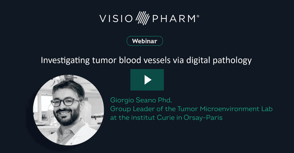
Dr Seano will present an overview of his works on blood vessel histological investigation. The study of the abnormalities of tumor blood vessels elucidated tumor features and is still shedding light on its pathology. Tumor vessels may be characterized by abnormal morphology, disrupted vascular basement membrane and reduced pericyte coverage. This causes dysfunction in perfusion and consequently leads to an hypoxic and immunosuppressive environment, typical of tumors. Breast cancer and glioblastoma are two of the most important tumors in the field of vascular microenvironment since we learnt that we can therapeutically modulate and temporally normalize vascular function. Dr Seano will show digital pathology results published during his period at Harvard Medical School and unpublished data from his own new lab.
Presented at the virtual Pathology Visions 2020 meeting during Visiopharm’s Industry Workshop on Monday, October 26, 2020.

Dr Giorgio Seano, Head of the Tumor Microenvironment LabInstitut Curie, Orsay-Paris (France)
Dr Seano is the Head of the Tumor Microenvironment Lab at Institut Curie, Orsay-Paris (France). His scientific interests are tumor vasculature, vessel co-option, cell migration and radioresistance. Among others, he published on Science, Cancer Cell, PNAS, Nat Cell Biol, Nat Biom Eng, JNCI, Blood and Cancer Res.