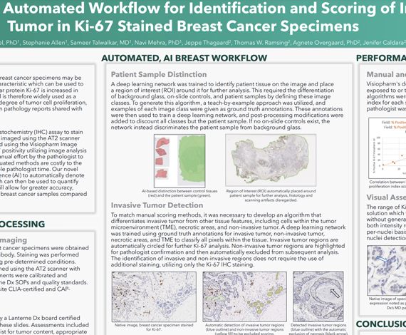
Understanding the rate of tumor cell growth in breast cancer specimens may be indicative of disease aggressiveness, a tumor characteristic which can be used to make an informed treatment decision. The nuclear protein Ki-67 is increased in cells as they prepare to divide, or proliferate, and is therefore widely used as a proliferation marker for tumor progression. This degree of tumor cell proliferation, or the proliferative index, is commonly detailed in pathology reports shared with the patient care team.
In this study, we utilized the Ki-67 [K2] immunohistochemistry (IHC) assay to stain 10 breast cancer specimens. Stained slides were imaged using the AT2 scanner (Leica Biosystems, Buffalo Grove, IL) and analyzed using the Visiopharm Image Analysis platform. Previous efforts to assess Ki-67 positivity utilizing image analysis have relied on the use of a secondary stain or manual effort by the pathologist to exclude non-invasive tumor regions. These antiquated methods are costly to the lab as they require additional materials or valuable pathologist time. Our novel image analysis approach utilizes artificial intelligence (AI) to automatically denote non-invasive verses invasive tumor regions, which can then be used to quantify the Ki-67 proliferative index. This valuable tool will allow for greater accuracy, cost-savings, and time efficiency when analyzing breast cancer samples compared to traditional methods.
Bhavika Patel, PhD1, Stephanie Allen1, Sameer Talwalkar, MD1, Navi Mehra, PhD1, Jeppe Thagaard2, Thomas W. Ramsing2, Agnete Overgaard, PhD2, Jenifer Caldara2
- Lanterne Dx, Boulder CO.
- Visiopharm Corporation