Transcript
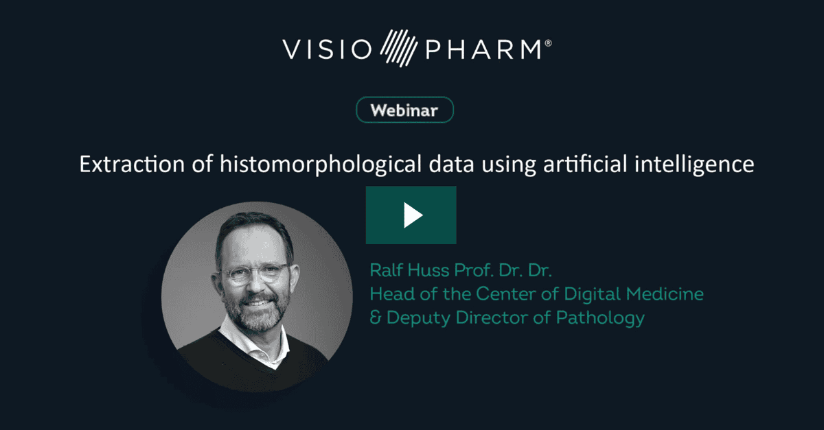
Hello and good afternoon, and welcome to Visiopharm’s session on the extraction of histomorphological data using artificial intelligence.
My name is Ralf Huss. I’m a pathologist and chairman of Visiopharm’s advisory board.
I would like to talk to you about why context still matters.
Context is in tissue that provides you spatial and qualitative information that is key to certain questions.
Let’s look at two different cases about the number on CD8 cells in tissue.
Both cases have the identical density.
However, they are unevenly distributed in the case number one where there are more cells in the stroma versus in the tumor.
While on the right side the number of CD8 cells are equally distributed between the tumor and the stroma.
And when you look at the distribution in the tissue, on the left side you might have the majority of CD8 cells outside of the tumor be in the stroma and not engaging with the tumor cells, while on the right side you have most CD8 cells directly inside the tumor, something that some of us would refer to as a hot tumor, versus a cold or excluded tumor on the left.
Currently, there are different opinions what is actually better for the patient.
So why is tissue still important while there are so many alternative methods that, however, do not show the full picture?
Tumor mutation burden, DAB, has its limitation.
For example, the tissue from non small cell lung cancer patients can or is usually very heterogeneous and is only a surrogate and therefore spatially resolved quantification performed by intelligent immunohistochemistry can overcome those challenges in making appropriate therapeutic decisions.
Therefore, the mutational density does not give you the whole picture because there is only a little correlation with the presence of infiltrating active T cells in the tumor.
Also liquid biopsies do not achieve the same sensitivity as whole tissue examination, although they can be drawn routinely and frequently from the same patient, which you usually cannot do with harvesting tissue from a patient.
So what is the unique information in intact tissue?
It is definitely the location of cells that matter.
Tissue resident and circulating peripheral immune cells are distinct population and have different tasks to fulfill.
Sometimes circulating immune cells first need to be primed before they can engage with the tumor in the tissue.
And also the spatial relationship between cell populations matter.
The context of cellularization is critical to understand the biology and the mode of action also of certain drugs.
And it is the tumor heterogeneity that matters, because TME is always heterogeneous and a modulator of tumor cell states.
Today’s workflow is a challenge and accordingly we face their limitations.
There is a demand for increasing biomarkers, which we sometimes try to cope with with immunofluorescence technologies.
Spatial information is crucial to determine cellular function when we try to identify the spatial relationships.
Increasing amount of data required for detailed analysis, therefore we try to add body fluid information. And all of that needs objective quantification to come up with a report to treat the patient with the best possible outcome.
Visiopharm’s solutions contribute to that. Their digital analysis overcomes those barriers.
Visiopharm offers high and ultraplex staining analysis, automated batch processing, tissue microenvironment analysis, standardized, validated and precise data analysis and all of the integration into data management systems.
Artificial intelligence is not new in medical applications. But why did it take so long to make it into practice?
Artificial intelligence is around for more than fifty years, but only until recently the computers have become available to apply robust AI applications to medical problems, whether it’s artificial intelligence by itself per se or machine learning or more recently, deep learning.
So what is the role of artificial intelligence in image analysis?
The more knowledge you have about a certain disease, a function of a biomarker or a drug, the more knowledge you can put into a classical image analysis.
That means you start with an image and apply manual features and manual rules to achieve an output.
But the more you have abstract data and less knowledge, you can still apply manual features, but let the system run automated routes like in machine learning and provides you new output that gives you more insights and more information into the content, for example, of biomarkers in tissue.
And if you know hardly anything but have more and abstract data, you can even apply automatic features and automated rules, something we call deep learning, applying artificial neural networks, or convolutional neural networks.
This is what some experts call the black box and ask for explainable AI to make the results plausible and understandable before we apply them to clinical practice.
All of that provides AI solution by Visiopharm.
What are typical solutions or questions that Visiopharm solutions contribute to digital image analysis?
Typical questions are, can I restrict analysis to invasive tumor only without manual annotations?
How can I quickly assess all cell populations in my sample?
What is the effect of my compounds in kidney and liver?
Has my compound reached the target in the tumor area?
What is the p d l one status in my tissue?
What are the different immune profiles of my cohort?
These are all different examples.
Let me give you some answers, For example, about the tumor detection.
With the appropriate app, you can have an automated tumor stroma separation and the detection of the tumor area where you can then quantify certain markers restricted to the tumor site.
Or how can I quickly assess all those different cell populations in my sample, which is too complex to do by human eye?
With the appropriate app, you can classify all those different cell types. You can put them in the right context, their spatial relationships, and come up with the appropriate heat map.
Or, for example, quantify p l one in lung cancer.
As you see here, detecting the tumor and quantifying PD L1 positive cells.
So Visiopharm provides a broad portfolio of AI based solutions that brings your research and your clinical practice forward. Whether you analyze tissue microarrays, you would like to build your own AI assisted apps, or you use one of the more than one hundred AI integrated ready to use apps, whether you wanna phenotype your multiplex analysis or you’d want to do virtual multiplex analysis.
These are just some examples of the broad portfolio that Visiopharm can provide to you on the basis of AI.
Thank you very much.
Data extraction from virtual slides is the foundation for the identification of novel patterns that are difficult or impossible to quantify by the human eye. Along with innovative technologies like high plex immunofluorescence, artificial intelligence allows the caption of deeper insights, contextual information, and describes spatial relationships.
Presented at the virtual Pathology Visions 2020 meeting during Visiopharm’s Industry Workshop on Monday, October 26, 2020.

Prof Ralf Huss, University Hospital Augsburg
Ralf Huss is a Professor of Pathology and the Managing Deputy Director of Pathology and Molecular Diagnostics at the University Hospital in Augsburg, Germany. He also heads the Center for Digital Medicine. He is certified in anatomical, experimental, and molecular pathology, with over 30 years of experience in histopathology, immunology, cancer research, and oncology.