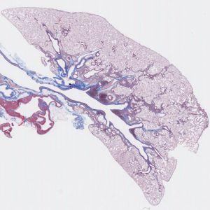
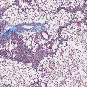
Rat lung stained with Masson’s trichrome stain.
#10040
Pulmonary fibrosis is the formation or development of excess fibrous connective tissue (fibrosis) in the lungs. Pulmonary fibrosis is a gradual exchange of normal lung parenchyma with fibrotic tissue, causing irreversible decrease in oxygen diffusion capacity. In addition, decreased compliance makes pulmonary fibrosis a restrictive lung disease. It is the main cause of restrictive lung disease that is intrinsic to the lung parenchyma. With Masson’s trichrome, the collagen in a rat lung section is stained blue and therefore very easy to distinguish from other tissue types. A magnification of 5X and a random 50% sampling is sufficient for an accurate and robust estimation of the collagen to tissue ratio.
Rat lung stained with Masson’s trichrome stain.
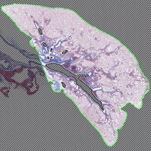
Specification of the region of interest. This can be performed automatically but requires manual inspection.
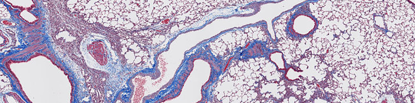
Close up of the lung tissue, consisting of blue stained collagen tissue and other tissue types.
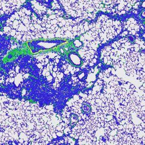
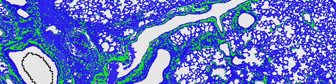
The final segmentation. The tissue is automatically segmented within a specified region of interest and divided into collagen tissue (green label) and other tissue (blue label).
Auxiliary APPs
Auxiliary APPs are used for additional process steps, e.g. finding Region of Interest (ROI).
ROI DETECT
This auxiliary APP ’01 ROI Detect’ can be used to automatically detect a ROI. The ROI is generated by distinguishing tissue from background.
Quantitative Output variables
The output variables of the analysis protocol includes:
Methods
The first image processing step involves a segmentation of the lung tissue from the background. This can be done automatically or manually depending on the complexity and quality of the image. As the collagen is quite distinct from the rest of the tissue, a threshold classifier is applied to a Red-Blue Contrast band. This segments the bluish collagen from the rest of the tissue.
Keywords
Pulmonary, Lung, Fibrosis, Collagen, quantitative, digital pathology, image analysis.