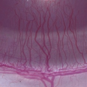
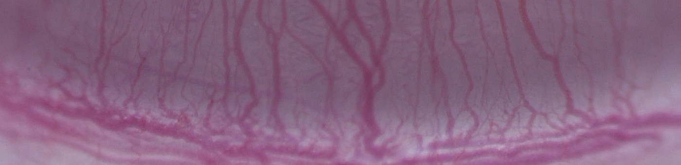
Brightfield image of the rat cornea, that shows the challenges associated with the segmentation of the vessels due to low color contrast.
#10021
The formation of new blood vessels by angiogenesis is an integral part of both normal developmental processes and numerous pathologies, ranging from tumor growth and metastasis to inflammation and ocular disease. Preclinical studies of disease mechanisms and the search for efficient treatments therefore includes assays for quantification of the effects of stimulators and inhibitors of angiogenesis. In certain diseases of the eye, angiogenesis is one of the fundamental processes where new blood vessels originating from the corneal veins will extend into the corneal stroma. This process, which is a biological consequence of inflammation, can also be used as an angiogenesis assay e.g., for testing agents with cancer treatment potential. The in vivo corneal neovascularization assay is based on the introduction of an angiogenic factor, such as vascular endothelial growth factor (VEGF) into the cornea of experimental animals, and then monitoring the effect of an inhibitor administered locally or systemically. The capillary growth, which takes place in a uniform direction towards the VEGF implant (see FIGURE 1), can then be monitored and quantified over time.
Brightfield image of the rat cornea, that shows the challenges associated with the segmentation of the vessels due to low color contrast.
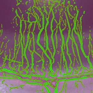
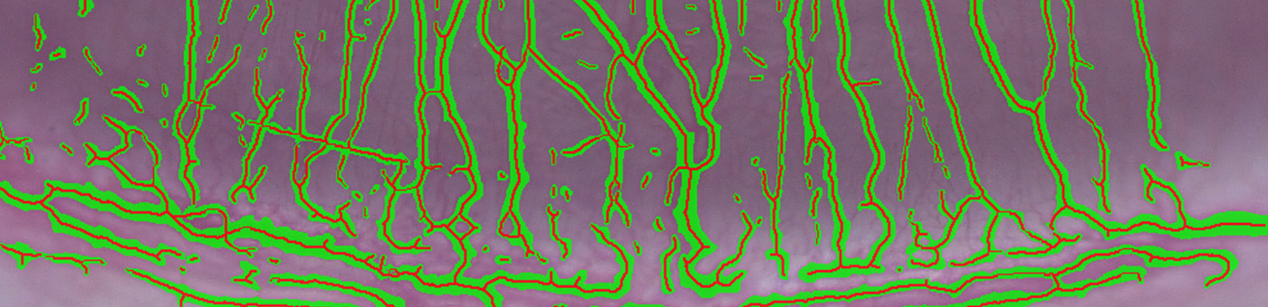
Segmentation of the vessel structure shown in green with the skeletonization super-imposed in red.
Quantitative Output variables
The output variables obtained from this protocol include:
Methods
Prior to the image analysis, it is important to outline the Region of Interest (ROI), thereby limiting the analysis from including baseline vessels and the vessels closest to the VEGF.
To enhance the vessels, while suppressing the background variation, several preprocessing steps are included. The more important steps include doing a median filtering to remove the variation of the background and applying a polynomial linear filter to enhance the blood vessels which have a very linear structure. The polynomial linear filter implementation is proprietary to Visiopharm and can effectively enhance most linear structures.
The classification identifies the entire vessel area within the region of interest. However, there is no straightforward way to translate the area for this complex structure into an estimate of length. Instead a skeletonization is applied after discarding all objects smaller than a (user-) defined limit. The skeletonize-step transforms the overlaying label to a 1-pixel wide structure by removing all pixels that do not belong to the midline (see FIGURE 2). From this, the total vessel length and individual segment lengths are calculated.
Keywords
Angiogenesis, bioassay, antiangiogenic, VEGF, quantitative, digital pathology, image analysis.
References
LITERATURE
1. Polverini, P.J., et. al. The Pathophysiology of Angiogenesis, Critical Reviews in Oral Biology and Medicine 1995, 6 (3), 230-47, DOI